Showing posts with label Palaeontologia Electronica. Show all posts

Saturday, December 6, 2025

[PaleoOrnithology • 2025] Chromeornis funkyi • A new small-bodied longipterygid (Aves: Enantiornithes) from the Aptian Jiufotang Formation preserving unusual gastroliths


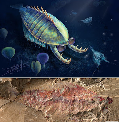
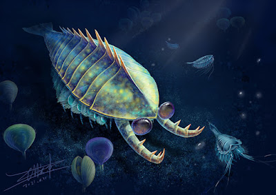
Chromeornis funkyi
O’Connor, Wang, Clark, Kuo, Davila, Wang, Zheng & Zhou, 2025 
 
Artwork: Sunny Dror
 
ABSTRACT
The Longipterygidae are a diverse group of small to medium sized enantiornithine birds with elongate rostra and distally restricted dentition known from the Early Cretaceous Jehol Lagerstätten. The largest taxon, Longipteryx, is known from dozens of specimens but comparatively little is known about small-bodied taxa, sometimes resolved in a subclade, the Longirostravinae. Here we describe a small longipterygid representing a new taxon, Chromeornis funkyi gen. et sp. nov., with a combination of features present in longirostravines and Longipteryx. Cladistic analysis indicates the new species is a member of the Longipteryginae, more closely related to Longipteryx than other longipterygids. The specimen preserves extensive soft tissue including traces of the eyes, skin, and feathers, as well as an unusual mass of gastroliths preserved appressed against the left lateral margin of the cervical vertebrae. Computed-tomography based comparison with the in situ gastric mill preserved in the sympatric ornithuromorphs Archaeorhynchus and Iteravis strongly suggests these gastroliths are not gizzard stones. The absence of a gastric mill in enantiornithines is consistent with pectoral girdle morphology that indicates limited flight capabilities in Early Cretaceous species suggesting ground take off, a necessity of collecting stones, was energetically costly compared to ornithuromorphs. Increases in body mass due to a large gastric mill may have further impeded volant locomotion resulting in a low cost-benefit tradeoff such that this structure was unlikely to evolve during early enantiornithine evolution.

 Keywords: Longipterygidae; new genus; new species; Jehol Biota; regurgitalite; gastrolith; Aves; Avialae.

Class AVES Linnaeus 1758
Clade ORNITHOTHORACES Chiappe 1995
Clade ENANTIORNITHES Walker 1981
Family LONGIPTERYGIDAE Zhang et al. 2000

Chromeornis funkyi gen. et sp. nov.
  
Etymology. Funky Chromeo bird, in honor of the Chromeo Funklordz P-Thugg and Dave 1, who like many birds, make beautiful music. Pronounced crow-me-OR-niss funk-e e.

Diagnosis. A small (estimated 33.5 g) longipterygid (rostrum ~60% of the skull or greater, distally restricted dentition, premaxillary corpus with elongate imperforate rostral end with parallel dorsal and ventral margins, robust pygostyle longer than tarsometatarsus, coracoid with straight lateral margin, humerus with narrow deltopectoral crest) enantiornithine (cranially forked pygostyle with ventrolateral processes, Y-shaped furcula with dorsally excavated rami, proximal humerus with small convex humeral head separated from the dorsal and ventral tubercles by concavities, minor metacarpal projecting farther distally than the major metacarpal, metatarsal IV reduced) distinguishable by the unique combination of the following characters: dentary straight; sternum with slightly splayed lateral trabeculae with asymmetrical fan-shaped distal expansions and short, straight intermediate trabeculae; hand shorter than humerus; alular digit short with small claw; second phalanx of major digit half the length of first phalanx; femur straight.


 Photographs of the counter slab of Chromeornis funkyi gen. et sp. nov. STM7-156.
 Scale bars equal one centimeter.
preserved with over 800 tiny rocks in its throat (visible as the gray mass next to the left of its neck bones). 

Close-up of the mass of rocks in the throat of Chromeornis (the rocks are the gray mass just to the left of the neck bones).  

An illustration showing Chromeornis funkyi gen. et sp. nov.  in life.
Artwork: Sunny Dror

  
Jingmai O’Connor, Xiaoli Wang, Alexander Clark, Pei-Chen Kuo, Ryan Davila, Yan Wang, Xiaoting Zheng, and Zhonghe Zhou. 2025. A new small-bodied longipterygid (Aves: Enantiornithes) from the Aptian Jiufotang Formation preserving unusual gastroliths. Palaeontologia Electronica. 28(3):a56. DOI: doi.org/10.26879/1589 
palaeo-electronica.org/content/2025/5712-longipterygid-enantiornithine-chromeornis
https://palaeo-electronica.org/content/current-in-press-articles/5713-longipterygid-enantiornithine-chromeornis


Saturday, October 18, 2025

[Paleontology • 2025] Oura megale • A large early Cambrian Deuteropod with a delta-shaped tailpiece


 Oura megale O’Flynn, Williams & Liu, 

in O’Flynn, Williams, Thomas, Hou et Liu, 2025. 

ABSTRACT
We describe Oura megale n. gen. n. sp., a large (ca. 14 cm long) euarthropod from the lower Cambrian Chengjiang Konservat-Lagerstätte of Yunnan Province, China. It possesses stalked, compound eyes and a possible raptorial frontal appendage (synapomorphies and symplesiomorphies of lower stem- and upper stem-group euarthropods, respectively), but in combination with deuteropodan synapomorphies (e.g., a multi-segmented head). Micro-X-ray fluorescence shows a thorax that consists of 10 comparatively long and five comparatively short segments. The terminal segment articulates with a large, delta-shaped tailpiece that may have conferred high positional manoeuvrability in the water column. The phylogenetic position of O. megale n. gen. n. sp. in the euarthropod stem may add support to the homology of raptorial frontal appendages between lower stem-group euarthropods and deuteropods.

Keywords: new genus; new species; Cambrian Chengjiang biota; euarthropod; micro-X-ray fluorescence; tailpiece

 Oura megale n. gen n. sp. (YKLP 17237).
A, photograph of ventral view. B, Fe map. C, composite line drawing. D, F1 map. E, P map. F, F1 map of head region. G, P map of head region.
 Abbreviations: l, left; r, right; A1, frontal appendage; ex.A6, exopod of 5th head limb on head segment 6; an, cephalic segment (n); as, anterior sclerite; cf, compactional fold; e, eye; es, eye stalk; exn, exopod (n); sn, sternite (n); Tn, thoracic exopod (n); tn, thoracic segment (n). Scale bars represent 20 mm.


SYSTEMATIC PALAEONTOLOGY

Phylum EUARTHROPODA Lankester, 1904

Unranked DEUTEROPODA Ortega-Hernández, 2016

Class, Order and Family incertae sedis

Genus OURA O’Flynn, Williams and Liu, new genus

Etymology. Gr. Oura, f. tail.

Remarks. Oura n. gen. is ascribed to Deuteropoda — as defined by a multi-segmented head and body arthrodization (Ortega-Hernández, 2016). Oura n. gen. bears: stalked, compound eyes, that is, a synapomorphy and symplesiomorphy of lower stem-group Euarthropoda and Deuteropoda, respectively (Ortega-Hernández, 2016, p. 11, table 4); and robust frontal appendages (FA, i.e., A1) that insert approximately one-half the length of the head behind the eye stalks and are inferred to be deutocerebral. The proximal-most three podomeres of the right-side frontal appendage (FA) are preserved, and these are morphologically like those of radiodonts and certain ‘great appendage’-bearing deuteropods.

Oura megale O’Flynn, Williams and Liu, new species
  
Etymology. Gr. Megale, large, great.

Diagnosis. Six cephalic segments. From anterior to posterior, these are: oblate spheroidal anterior sclerite to which large ovate paired stalked lateral eyes are attached; post-ocular segment with paired uniramous deutocerebral FAs; and four subsequent segments. FA antero-laterally orientated, consisting of ≥ three podomeres that bear elongate triangular spines. Thorax consists of 15 segments (anterior-most 10 comparatively long; posterior-most five comparatively short), each with one pair of presumed biramous appendages. Segment 15 articulated with large, ogival delta tailpiece.


Robert J. O’Flynn, Mark Williams, Ed Thomas, Xianguang Hou, and Yu Liu. 2025. Oura megale n. gen. n. sp., A large early Cambrian Deuteropod with a delta-shaped tailpiece. Palaeontologia Electronica. 28(3):a44. DOI: doi.org/10.26879/1547 
palaeo-electronica.org/content/2025/5687-early-cambrian-deuteropod-oura-megale
 x.com/PalaeoE/status/1977837034074210364

Plain Language Abstract:  Oura megale, a large arthropod (segmented, jointed-limbed animal), comparable in size to anomalocaridids (i.e., giant invertebrate Cambrian predators featuring mouthparts composed of a ring of plates), inhabited the Cambrian seas (approximately 518 million years ago) of south-western China. It had stalked eyes and a grasping frontal appendage, but in combination with a multi-segmented head, as we see in, for example, modern insects. Palaeontological techniques show a trunk that consists of 10 long and five short segments. The rearmost segment is associated with a large, delta-shaped tail that may have been conducive to efficient swimming. The evolutionary relationships between O. megale and other arthropods may suggest that the grasping appendage (shared by several arthropods) is inherited from a common ancestor.

Friday, August 22, 2025

[PaleoMammalogy • 2025] Megabalaena sapporoensis • A New member of a large and archaic balaenid (Mysticeti: Balaenidae) from the Late Miocene of Sapporo, Hokkaido, Japan partly fills a gap of Right Whale Evolution


Megabalaena sapporoensis
Tanaka, Kimura, Shinmura, Ohira & Furusawa, 2025

 Artwork by Tatsuya Shinmura 

ABSTRACT
The family Balaenidae (right whales) includes two genera and four extant species, all of which are endangered and giant animals measuring approximately 17 to 20 m in length. The history of the Balaenidae spans about 20 million years. Several small sized extinct balaenids from the Pliocene have been identified. However, half of this history remains unknown owing to a 9-million-year gap from 15.2 to 6.1 m.y.a. in the fossil record. A well-preserved fossil balaenid skeleton, designated SMAC 2731, from the Late Miocene approximately 9 m.y.a. in Sapporo, Hokkaido, Japan, is named as Megabalaena sapporoensis gen. et sp. nov. This specimen preserves the skull, periotics in situ, tympanic bullae, right mandible, basihyal-thyrohyal, right stylohyal, sternum, seven cervical vertebrae, nine thoracic vertebrae, and 16 more posterior vertebrae, rib fragments, scapulae, and left forelimb elements. All preserved vertebral epiphyses are fused, indicating that SMAC 2731 was physically mature. Notably, M. sapporoensis can be distinguished from other balaenids by its excavated orbit in dorsal view with a large postorbital process, dorsoventrally high anterior part of the involucrum of the tympanic bulla, long compound posterior process, high coronoid process and deeper subcondylar furrow of the mandible, incipient cervical fusion (C2+C3 only), and its slender forelimb bones, including the humerus, radius and ulna. Based on a bizygomatic width of 2.2 m, the estimated total length of the holotype of M. sapporoensis is 12.7 m. Overall, M. sapporoensis indicates that balaenids diversified prior to the Late Miocene.
   
Keywords: Balaenidae;  new genus;  new species;  Tortonian;  gigantism;  Japan

Forelimb elements of balaends. 
Megabalaena sapporoensis (A),
Charadrobalaena valentinae, outline taken from Bisconti et al. (2023) and is a mirror image (B),
Antwerpibalaena liberatlas, outline taken from Duboys de Lavigerie et al. (2020) and is a mirror image (C),
Eubalaena japonica, outline taken from Omura (1958) (D), and are not to scale.

  Images based on a 3D model showing preserved skull elements of SMAC 2731, Megabalaena sapporoensis. Deformations are not restored.

 Images based on a 3D model showing skull elements of SMAC 2731, Megabalaena sapporoensis in left lateral view (A) and dorsal view (B). Deformations are restored using 3D model editor by T. Shinmura. Settings are the same to Figure 3.

CETACEA Brisson, 1762
NEOCETI Fordyce and de Muizon, 2001

MYSTICETI Gray, 1864
CHAEOMYSTICETI Mitchell, 1989

BALAENIDAE Gray, 1825

Megabalaena gen. nov 
Type species. Megabalaena sapporoensis sp. nov.

Etymology. The generic name, Megabalaena , is named derived from ancient Greek megas meaning greatlarge and mighty, and the type genus name of the family Balaenidae.
 
Megabalaena sapporoensis sp. nov.

Locality and horizon. SMAC 2731 was found at a riverbed of Toyohira River in Sapporo City, Hokkaido, Japan, by Kazuhisa Mori on 10 October 2008: Latitude 42°58'1.24"N, longitude 141°13'18.01"E (Figure 1 and Figure 2). SMAC 2731 was found from the upper part of the Toyama Formation. At the fossil area, the diatomaceous siltstone Toyama Formation is distributed. Diatomaceous siltstone of the Toyama Formation is exposed at the type locality ...

Etymology. Named after the fossil locality, Sapporo City.

Diagnosis. Megabalaena sapporoensis is a member of the Balaenidae because it has a combination of these character states such as a posteriorly pointed anterior edge of the supraorbital process lateral to the ascending process of the maxilla with the skull in dorsal view (Character 31, state 0), laterally oriented postorbital process in dorsal view (Character 38, state 1), confluent posterior border of the zygomatic process of the squamosal and exoccipital in dorsal view (Character 67, state 1), dorsoventrally higher than long parietal in lateral view (Character 76, state 1), anterolaterally directed zygomatic process of the squamosal in dorsal view (Character 86, state 2), distinctly higher than long squamosal including the zygomatic and postglenoid processes (Character 92, state 1), short squamosal fossa (Character 96, state 1), foramen pseudovale opening posteriorly between the squamosal and pterygoid (Character 118, state 1), and posteriorly diverging basioccipital crests in ventral view (Character 125, state 0).
..

 Restoration of Megabalaena sapporoensis by Tatsuya Shinmura (Ashoro Museum of Paleontology).


Yoshihiro Tanaka, Toshiyuki Kimura, Tatsuya Shinmura, Hiroto Ohira, and Hitoshi Furusawa. 2025. A New member of a large and archaic balaenid from the Late Miocene of Sapporo, Hokkaido, Japan partly fills a gap of Right Whale Evolution.  Palaeontologia Electronica. 28(2): a37. DOI: doi.org/10.26879/1549 [August 2025]

  
Plain Language Abstract: The right whale family (Balaenidae) includes four extant species in two genera such as the Balaena and Eubalaena, all of which are endangered and giant animals about 17 to 20 m in length. The history of the right whale group spans about 20 million years. Several small-sized extinct fossil right whales from the Pliocene have been identified. However, half of this history remains unknown owing to a 9-million-year gap from 15.2 to 6.1 million years ago in the fossil record. A well-preserved fossil right whale skeleton (SMAC 2731) from the late Miocene (approximately 9 m.y.a.) of Sapporo, Hokkaido, Japan, is named as the new species Megabalaena sapporoensis. This specimen preserves the skull, ear bones, right lower jaw, hyoid bones, sternum, back bones, ribs, scapulae, and left forelimb elements. All preserved vertebral epiphyses are fused, indicating that SMAC 2731 was physically mature. Notably, M. sapporoensis can be distinguished from other balaenids by its large postorbital process of the skull and unfused cervical vertebrae, except for the axis and third cervical vertebra, and its slender forelimb long bones, including the humerus, radius and ulna, which are about twice slenderer than these of extant balaenids. Based on a bizygomatic width of 2.2 m, the estimated total length of the holotype of M. sapporoensis is 12.7 m. Overall, M. sapporoensis enhances our understanding of balaenid diversity, suggesting that it expanded earlier than the late Miocene.

Wednesday, October 9, 2024

[Paleontology • 2024] Ardetosaurus viator • A New diplodocine Sauropod from the Morrison Formation, Wyoming, USA


 Ardetosaurus viator
van der Linden, Tschopp, Sookias, Wallaard, Holwerda & Schulp, 2024

 DOI: 10.26879/1380 
  Life reconstruction by Ole Zant. x.com/TheBioBob

ABSTRACT
The Morrison Formation of the western United States is well-known for its high diversity of sauropod dinosaurs. The Howe-Stephens Quarry in northern Wyoming is one of several quarries which has yielded several associated to completely articulated dinosaur specimens, among which a semi-articulated diplodocid specimen, MAB011899, which was excavated in 1993. This diplodocid specimen is represented by posterior cervical, dorsal, sacral, and anterior caudal vertebrae, multiple thoracic ribs, two chevrons, a left coracoid, a left ilium, both pubes and ischia, a left femur, a left tibia, and a left fibula. Through comparative anatomy, we interpret this specimen as a new species of diplodocine sauropod, Ardetosaurus viator gen. et sp. nov. Unambiguous autapomorphies include paired accessory laminae in the spinoprezygapophyseal fossae of posterior cervical and anterior dorsal vertebrae, bifurcating anterior centrodiapophyseal laminae in the anterior dorsal vertebrae, fossae present in the centropostzygapophyseal laminae of the second dorsal vertebra, a low vertebral height/centrum length ratio of the posterior dorsal vertebrae and reduced to absent centroprezygapophyseal laminae in the anterior caudal vertebrae. Local autapomorphic features include single centroprezygapophyseal laminae in the posterior cervical vertebrae and a highly elliptical cross-section of the femoral midshaft. Ardetosaurus viator is the first skeletally mature sauropod specimen described from the Howe-Stephens Quarry. This specimen provides insight into serial variation of vertebral laminae and laminar transitions. Finally, the peculiar morphology of the—often not preserved—first chevron is described in detail, and its possible use in studying sexual dimorphism in sauropods is discussed.

Keywords: sauropod; new genus; new species; Morrison Formation; Diplodocinae; Wyoming


  Skeletal reconstruction of  Ardetosaurus viator MAB011899. Skeletal reconstruction indicating preserved bones (white), excavated bones but subsequently lost (light gray) and not preserved (dark gray). Unknown elements are based on other diplodocines.
Scale bar equals 1 m. Reconstruction by Ole Zant.

SYSTEMATIC PALEONTOLOGY
DINOSAURIA Owen, 1842
SAUROPODA Marsh, 1878
EUSAUROPODA Upchurch, 1995
NEOSAUROPODA Bonaparte, 1986

DIPLODOCOIDEA Marsh, 1884
FLAGELLICAUDATA Harris and Dodson, 2004

DIPLODOCIDAE Marsh, 1884
DIPLODOCINAE Marsh, 1884

ARDETOSAURUS gen. nov.
 
Ardetosaurus viator gen. et sp. nov.
 
Holotype. MAB011899: two cervical vertebrae, 10 dorsal vertebrae, sacrum, five caudal vertebrae, eight dorsal ribs, two chevrons, a left coracoid, a left ilium, both pubes, both ischia, a left femur, a left tibia, and a partial left fibula.

Diagnosis. Ardetosaurus viator is diagnosed by the combination of the following autapomorphies: 1) the presence of distinct, paired accessory laminae in the spinoprezygapophyseal fossae (SPRF) in the posterior cervical and anterior dorsal vertebrae, 2) anteroventrally bifurcating anterior centrodiapophyseal laminae (ACDLs) in the anterior dorsal vertebrae, 3) the presence of centropostzygapophyseal lamina fossae (CPOL-f) in the second dorsal vertebra, 4) a vertebral height/centrum length ratio of <2.5 of the posterior dorsal vertebrae, and 5) reduced or absent centroprezygapophyseal laminae (CPRLs) in the anterior-most caudal vertebrae. Ardetosaurus viator differs from all other diplodocines by having unbifurcated CPRLs in the posterior cervical vertebrae and a highly elliptical femoral cross-section. Ardetosaurus viator differs from Amphicoelias Cope, 1878, in lacking the rounded, lateral projections of the neural spine tip and the thin neural spine base in the dorsal vertebrae; from Barosaurus Marsh, 1890, by having tall cervical neural spines, single midline keels, narrower prezygapophyseal rami in the cervical vertebrae, ten dorsal vertebrae, the presence of ...


Etymology. ‘Ardeto’ is an inflection of Latin ārdēre, meaning ‘to burn.’ It refers to the history of some of the elements, which were either fully destroyed in a fire, or still show burn scars from the fire. ‘saurus, ’ Latinized form of the Greek σαῦρος (saúros), meaning lizard or reptile. ‘viator’ is Latin for traveler, referring to the journey of the specimen from the USA, via Switzerland and Germany, to the Netherlands.

Locality and horizon. Ardetosaurus viator comes from the Howe-Stephens Quarry of northern Wyoming, USA. The quarry is dated, based on magnetostratigraphy and correlation with other sections in the Morrison basin (Maidment and Muxworthy, 2019; Maidment, personal communication, 2022) at 150.44 to 149.21 million years old, placing it in the Kimmeridgian Stage of the Upper Jurassic.


 Quarry map of Ardetosaurus viator MAB011899. Excavation map of the Howe-Stephens Quarry, indicating the major finds from 1992-2000. Individual dinosaurs are color coded, and MAB011899 is coded with dark blue, and named ‘Diplodocus Brösmeli’ herein. The red crosses indicate the missing/lost cervical vertebrae. Note the relatively similar color for ‘Brösmeli’ and ‘David’ (SMA 0086), but their significant separation in the quarry.
 Figure is courtesy of the SMA. Quarry sections equal 1 by 0.5 m.


  Life reconstruction of  Ardetosaurus viator MAB011899.
Illustration by Ole Zant.

The skeleton of Brösmeli is on display in the Oertijdmuseum.


Tom T.P. van der Linden, Emanuel Tschopp, Roland B. Sookias, Jonathan J.W. Wallaard, Femke M. Holwerda, and Anne S. Schulp. 2024. A New diplodocine Sauropod from the Morrison Formation, Wyoming, USA. Palaeontologia Electronica. 27(3): a50. DOI: doi.org/10.26879/1380
palaeo-electronica.org/content/2024/5327-new-diplodocine-sauropod

Wednesday, July 24, 2024

[Paleontology • 2024] Propterodacylus frankerlae • A pterosaurian connecting link from the Late Jurassic of Germany

 
 Propterodacylus frankerlae
Spindler, 2024


ABSTRACT
Based on a unique and extraordinarily preserved complete skeleton, the “Painten pro-pterodactyloid” is formally described and named as Propterodactylus frankerlae, gen. nov., spec. nov. As previously shown, it has a nearly perfect mix of plesiomorphic rhamphorhynchoid-grade, wukongopterid, and derived pterodactyloid traits. Due to its lack of autapomorphies, Propterodactylus is a sufficient intermediate taxon that closes the greatest knowledge gap regarding the evolution of pterosaur morphology. Non-pterodactyloid features include interlocking caudal vertebrae and a functional fifth pedal toe. Derived features such as the nasoantorbital fenestra, a short tail, or initially elongated cervicals and metacarpals appear ancient within the spectrum of Pterodactyloidea. Other early Monofenestrata appear more autapomorphic. However, the late juvenile or subadult status of the described specimen suggests that the rostrum, neck, and extremities might have been even more elongated when fully grown. Despite the otherwise intermediate, transitional osteology, details of dentition types appear mosaic-like in early Monofenestrata. While the remainder of the skeleton of Propterodactylus fits into known evolutionary trends, its dentition implies a significant role of varying dietary adaptation throughout the pterodactyl transition.

Keywords: new genus; new species; Pterosauria; evolution; dentition

 Specimen DMA-JP-2011/006, holotype of Propterodacylus frankerlae, gen. nov., sp. nov.
(A) under daylight condition; (B) under UV (365 nm),
by courtesy of Helmut Tischlinger. Scale bars equal 5 cm.

SYSTEMATIC PALAEONTOLOGY

Order Pterosauria Owen, 1842 (for a rejection of Pterosaurii Kaup, 1834 see Seeley, 1870, 1891)

Clade Monofenestrata Lü, Unwin, Jin, Liu, and Ji, 2010

Propterodactylus gen. nov. 

Remarks. The intermediate status is underlined by a lack of apparent autapomorphies. Tischlinger and Frey (2013) listed several plesiomorphic and apomorphic traits. Similarities with the derived Pterodactyloidea comprise the skull shape and short tail. Plesiomorphies shared with e.g., Wukongopteridae, which preclude Propterodactlyus from Pterodactyloidea, are the functional fifth toe and long caudal zygapophyses. Intermediate conditions apply to the cervical elongation, metacarpal elongation, and reduced fifth toe.

Etymology. The genus name refers to the informal designation as a pro-pterodactyloid in the literature, meaning a supposed forerunner (ancient Greek προ- for “before”) of the iconic Pterodactylus (latinized form of Greek πτερόν plus δάκτυλος for “wing digit”) and at the same time a forerunner of the Pterodactyloidea in general.


 Propterodactylus frankerlae, gen. nov., spec. nov. 

Etymology. The epithet honours Petra Hahn, née Frankerl (1966 - 2019), the wife of Stephan Hahn, who found the specimen in 2011 during the scientific excavation.


Frederik Spindler. 2024. A pterosaurian connecting link from the Late Jurassic of Germany. Palaeontologia Electronica. 27(2):a35. DOI: 10.26879/1366

Tuesday, April 16, 2024

[PaleoIchthyology • 2024] Toarcocephalus morlok • First Occurrence of A coccolepidid fish (?Chondrostei: Coccolepididae) from the Upper Lias (Toarcian, Early Jurassic) of southern Germany


Toarcocephalus morlok 
Cooper,  López-Arbarello & Maxwell, 2024

Artwork by S. Cooper.

ABSTRACT
The non-neopterygian group †Coccolepididae, a moderately diverse predominantly freshwater family, remains an imperfectly known Mesozoic group of actinopterygians, currently classified within Chondrostei based on the presence of several acipenseriform synapomorphies. Coccolepidids first appear during the Early Jurassic in marine sediments, although their fossils are poorly known from this time, and none have yet been described from the Toarcian (Upper Lias). Here, we describe a new genus and species of coccolepidid fish, †Toarcocephalus morlok gen. et sp. nov. from the Lower Jurassic (Toarcian) Posidonienschiefer Formation of Holzmaden in southern Germany. †Toarcocephalus morlok is diagnosed by a unique combination of characters including a shallow lower jaw with a massive angular, opercle and subopercle equal in size, preopercle that only borders the subopercle but does not reach the opercle; dermal skull bones strongly punctate, with externally smooth upper and lower jaw bones. Discovery of a coccolepidid at Holzmaden represents the first occurrence of the group from a Toarcian deposit, as well as the oldest record of the family in mainland Europe. Both described specimens of †T. morlok were victims of successful predation events: one individual was likely decapitated (pabulite) and the other preserved within a regurgitalite (fossilized oral ejecta). The evolution of Coccolepididae is discussed briefly in relation to a marine/freshwater origin.

Keywords: Coccolepididae; Chondrostei; Posidonienschiefer Formation; Early Jurassic; paleobiogeography; regurgitalite

 Cranial reconstruction of †Toarcocephalus morlok gen. et sp. nov. with missing regions based on reconstruction of ‘Coccolepis’ liassica in Gardiner (1960). Missing or speculative areas are indicated with a dashed line and coloured in grey.
 Abbreviations: ag = angular; an = antorbital; br = branchiostegal rays; cl = cleithrum; d = dentary; dpt = dermopterotic; dsph = dermosphenotic; esc = extrascapular; g = gular plate; hyo = hyomandibula; io.c = infraorbital sensory canal; j = jugal; msc = mandibular sensory canal; mx = maxilla; na = nasal; nar = external naris; op = opercle; poc = postorbital canal; pop = preopercle; ro = rostral; scl = supracleithrum; scr = sclerotic ring; sop= subopercle.


SYSTEMATIC PALAEONTOLOGY 
Class ACTINOPTERYGII Cope, 1887
Subclass ?CHONDROSTEI Müller, 1845 sensu Grande and Bemis (1996)
Family †COCCOLEPIDIDAE Berg, 1940 sensu López-Arbarello et al. (2013)

Toarcocephalus gen. nov.

Toarcocephalus morlok gen. et sp. nov.
 
Diagnosis. †Toarcocephalus morlok gen. et sp. nov. is diagnosed from all other coccolepidid fishes by the following unique combination of characters: upper and lower jaws smooth and unornamented; mandible well elongated and gracile, longer than maxilla and shallow posteriorly; angular large and lenticular; large postorbital expansion of maxilla strongly convex and twice as long as deep with a strongly recurved ventral margin; short premaxilla holding several recurved teeth that are slightly larger than those on the maxilla; skull roof very weakly tuberculated with pronounced striated ridges that are marginally serrated; subopercle trapezoidal and equal in size to opercle; preopercle slender, forming a posteroventral lobular expansion, only as tall as the subopercle and extending no further than the midpoint of the postorbital plate of the maxilla; operculum and supracleithrum mostly smooth with fine, regularly spaced punctae; supracleithrum massive, accounting for more than 80% of operculum height; large triangular dorsal process on the supracleithrum; nine branchiostegal rays, each thin, lacking distal expansions and confined to the posterior corner of the mandible; gular plate egg-shaped and placed roughly at the midpoint of the lower jaw length; elongated hyomandibula obliquely inclined forward, thin and weakly bow-shaped; ceratohyal well elongated but thin; hypohyals short and robust; sclerotic ring thin and delicate; scales weakly developed; vertebral column aspondylous, composed of simple arcocentral arches with proportionately short spines.

Etymology. Generic name chosen for its discovery in Toarcian-aged strata, with suffix -cephalus for head, denoting the diagnostic cranium. Species epithet morlok named after the savage subterranean antagonists in H.G. Wells’ The Time Machine (1895), due to their similarly grotesque appearance characterised by large eyes, a blunt face and pointed teeth.

 Palaeoart depiction of †Toarcocephalus morlok gen. et sp. nov. swimming in the twilight of the Posidonia Shale Sea.
The post-cranium is based on †‘Coccolepisliassica and †Coccolepis bucklandi.
Artwork by S. Cooper.


Samuel L.A. Cooper,  Adriana López-Arbarello and Erin E. Maxwell. 2024. First Occurrence of A †coccolepidid fish (?Chondrostei: †Coccolepididae) from the Upper Lias (Toarcian, Early Jurassic) of southern Germany. Palaeontologia Electronica. 27(1):a23. DOI: 10.26879/1326  

Friday, February 2, 2024

[Paleontology • 2024] Ophiussasuchus paimogonectes • A New goniopholidid Crocodylomorph (Crocodylomorpha: Goniopholididae) from the Late Jurassic of Portugal


Ophiussasuchus paimogonectes
López-Rojas, Mateus, Marinheiro, Mateus, & Puértolas-Pascual, 2024


ABSTRACT
Plenty of goniopholidid species from the Mesozoic have been found in the Iberian Peninsula. A previous goniopholidid taxon, Goniopholis baryglyphaeus Schwarz, 2002, from the Late Jurassic of the Guimarota coal mine (Leiria, central Portugal) was described. This taxon corresponds to a partial skull and some postcranial material, and it marked the oldest and first ever record of goniopholidid and Goniopholis species described for the Iberian Peninsula. Here we present a well-preserved, almost complete, skull of a new species, Ophiussasuchus paimogonectes gen. et sp. nov. from Upper Jurassic deposits of Praia de Paimogo (near Lourinhã, central west Portugal). The specimen corresponds to a mesorostrine, platyrostral skull of a medium-sized goniopholidid coming from the upper Kimmeridgian within the Lourinhã Formation. Phylogenetically, the new species is recovered as the sister taxon of the Early Cretaceous European clade made by Hulkepholis and Anteophthalmosuchus. Although its position is well-resolved, this new taxon displays intermediate morphological traits, sharing characteristics with Jurassic Asian and American basal goniopholidids (e.g., presence but lesser development of the secondary choana with the nasopharyngeal duct partially open), as well as more derived characters shared with Cretaceous European taxa such as Hulkepholis (e.g., the shape of the supratemporal fenestra and the palatines). As a result, Ophiussasuchus paimogonectes gen. et sp. nov. exhibits characteristics suggesting a reversion to primitive goniopholidid conditions or intermediate states between the goniopholidid taxa of North America and Europe. These findings support the shared Late Jurassic fauna between the Morrison and Lourinhã Formations, while also having high endemism of taxa.

Keywords: new genus; new species; Lourinhã Formation; upper Kimmeridgian; Crocodylomorpha; phylogeny

 Skull of ML2776 Ophiussasuchus paimogonectes gen. et sp. nov. 
Anatomical abbreviations: f, frontal; itf, infratemporal fenestra; j, jugal; lac, lacrimal; mx, maxilla; mx.dp, maxillary depressions; n, nasal; o, orbit; pa, parietal; pal, palpebral; pf, prefrontal; pmx, premaxilla; po, postorbital; q, quadrate; qj, quadratojugal; sq, squamosal; stf, supratemporal fenestra; t, tooth. 
Scale represents 10 cm.

 Skull of ML2776 Ophiussasuchus paimogonectes gen. et sp. nov., in lateral view.
Anatomical abbreviations: ec, ectopterygoid; j, jugal; mx, maxilla; mx.dp, maxillary depressions; n, nasal; pal, palpebral; pf, prefrontal; pmx, premaxilla; po, postorbital; pt, pterygoid; q, quadrate; qj, quadratojugal; sq, squamosal; t, tooth. 
Scale represents 10 cm.

SYSTEMATIC PALEONTOLOGY

Superorder CROCODYLOMORPHA Hay, 1930 (sensu Walker, 1970)
Clade CROCODYLIFORMES Hay, 1930

Suborder MESOEUCROCODYLIA Whetstone and Whybrow, 1983 (sensu Benton and Clark, 1988)
Infraoder NEOSUCHIA Gervais, 1871 (sensu Benton and Clark, 1988)

Family GONIOPHOLIDIDAE Cope, 1875

Genus OPHIUSSASUCHUS gen. nov.

Etymology. ‘ Ophiussa ’ refers to the ancient name given by the Greeks to the area where is now Portugal; and ‘ suchus ’ is from the Greek souchos that refers to crocodile-headed Egyptian god Sobek.

OPHIUSSASUCHUS PAIMOGONECTES sp. nov
 
Holotype. ML2776, an almost complete and well-preserved isolated skull, with some teeth still attached. The specimen is deposited at Lourinhã Museum (Museu da Lourinhã), Lourinhã, Portugal.

Etymology. ‘paimogonectes’ refers to the one who swims in Paimogo, where the specimen was found (Paimogo beach, Lourinhã, Portugal).

Age and horizon. Praia Azul Member, Lourinhã Formation, Lusitanian Basin, west coast of Portugal. upper Kimmeridgian-lower Tithonian, Upper Jurassic.

Type locality. Praia de Paimogo (GPS coordinates: 39° 17’ 10.4” N, 9° 20 ’17.4” W), Lourinhã, Lisbon, Portugal.

Diagnosis. Medium-sized crocodylomorph about 2.5 m to 3 m long (estimated body length based on Young et al., 2011), with platyrostral, mesorostrine skull and a marked festooned contour which differs from other goniopholidids since it possess a less pronounced axe-shaped premaxillae dorsal outline; smooth perinarial region with absence of crests around it; presence of different number of maxillary depressions between the left and right regions (four and five, respectively); anteriorly well-marked V-shape border of the palatines in contact with the maxillae; nasals with straight, sub-parallel margins between the maxillae, with little to no lateral expansion at their posterior-most border; main body of the frontal with sub-squared shape in dorsal view, without lateral expansion; nasopharyngeal duct less ventrally exposed than in Jurassic taxa (e.g., Calsoyasuchus or Eutretauranosuchus) but not as closed as in Cretaceous taxa (e.g., Hulkepholis or Anteophthalmosuchus); presence of two small, thin, anteroposteriorly elongated and crescent-shaped palatal fenestrae between the maxillae and the palatines.


CONCLUSIONS: 
A new and the most complete skull of crocodylomorph (ML2776) from the Late Jurassic of Lourinhã Fm, was recovered and is here described as a new genus and species, Ophiussasuchus paimogonectes gen. et sp. nov.

This new taxon was recovered with a unique combination of 15 synapomorphies. These synapomorphies are a combination of features from both older North American and younger European taxa, such as the shape of the rostrum with a less-projected axe shape compared with Jurassic North American species (Amphicotylus) and more similar to Cretaceous taxa, while the nasopharyngeal duct openings are more closed than older species (Calsoyasuchus or Eutretauranosuchus) but not completely closed as in Hulkepholis and Anteophthalmosuchus, with which Ophiussasuchus paimogonectes gen. et sp. nov. has closer relationships. In addition, ML2776 displays an asymmetrical number of maxillary fossae (four in the left region while five in the right region). However, any hypothesis regarding this asymmetry cannot be contrasted until more specimens are found.

The new taxon Ophiussasuchus paimogonectes gen. et sp. nov. increases the already high paleodiversity of crocodylomorphs of the locality. It also supports the hypothesis of a shared fauna between North America (Morrison Fm) and Western Europe (Lourinhã Fm) during the Late Jurassic, as well as the high endemism of Portugal, distinguishing both continents.

 
Víctor López-Rojas, Simão Mateus, João Marinheiro, Octávio Mateus, and Eduardo Puértolas-Pascual. 2024. A New goniopholidid Crocodylomorph from the Late Jurassic of Portugal. Palaeontologia Electronica, 27(1):a5. DOI: 10.26879/1316


Thursday, May 11, 2023

[Paleontology • 2023] A Reappraisal of the Cranial and Mandibular Osteology of the spinosaurid Irritator challengeri (Dinosauria: Theropoda)


Irritator challengeri  Martill, Cruickshank, Frey, Small & Clarke, 1996

in Schade, Rauhut, Foth, Moleman et Evers, 2023. 

ABSTRACT
Although originally described almost three decades ago, the holotype of Irritator challengeri from the Lower Cretaceous Romualdo Formation of Brazil still represents the most complete spinosaurid skull known to science. Here, we present a detailed description of the skull of Irritator based on digital reconstructions from medical and micro computed tomography (µCT) data. Segmentation reveals the near-complete palatal complex and braincase, an unusual morphology of the retroarticular process, a large, ventrally inclined surangular shelf and the tooth replacement pattern. The digitally reconstructed skull anatomy indicates a robust dentition, a field of binocular vision in front of the skull with an inclined snout orientation, a relatively weak but fast bite, as well as laterally spreading and rotating lower jaw rami during jaw opening. We modified an existing phylogenetic matrix of Tetanurae to account for new observations on the morphology of Irritator and analysed this using parsimony and Bayesian methods. Results support Spinosauridae as members of Megalosauroidea and recover a monophyletic Carnosauria (Megalosauroidea + Allosauroidea). Parsimony analysis recovers Monolophosaurus nested within Megalosauroidea as sister taxon to spinosaurids, but this is not supported by the Bayesian analysis. Bayesian time-calibration and evolutionary rate analysis indicate that spinosaurid evolution happened fast, despite a long ghost lineage of at least 35 million years. High evolutionary rates over a prolonged time can explain the highly derived skull morphology of spinosaurids. This study provides an in-depth look into the evolution of spinosaurid skull anatomy and refines our understanding of these specialized Mesozoic predators.

Keywords: Dinosaur; Theropod; Spinosaurid; Cretaceous; Mesozoic; Brazil


Theropoda Marsh, 1881
Tetanurae Gauthier, 1986
Megalosauroidea Fitzinger, 1843; sensu Carrano et al. (2012)

Spinosauridae Stromer, 1915
Spinosaurinae (Stromer, 1915); sensu Sereno et al. (1998)

Irritator challengeri Martill, Cruickshank, Frey, Small and Clarke, 1996

Holotype. SMNS 58022, largely complete skull, missing most of the premaxillae, anterior ends of the maxillae, and anteriormost parts of both mandibles.

Locality and horizon. Near Buxexé, close to Santana do Cariri, Ceará State, northeastern Brazil (see Sues et al., 2002: 535). Lower part of the Romualdo Formation (Santana Formation of some authors; see discussion in Arai and Assine, 2020) of the Santana Group, late Aptian (Arai and Assine, 2020).


Marco Schade, Oliver W. M. Rauhut, Christian Foth, Olof Moleman, and Serjoscha W. Evers. 2023. A Reappraisal of the Cranial and Mandibular Osteology of the spinosaurid Irritator challengeri (Dinosauria: Theropoda). Palaeontologia Electronica. 26(2): a17.DOI: 10.26879/1242
 
palaeo-electronica.org/content/2023/3821-the-osteology-of-irritator


Friday, March 3, 2023

[Paleontology • 2022] Fengzhengia mamingae • A New Euarthropod with Large frontal Appendages from the early Cambrian Chengjiang Biota, Chengjiang Lagerstätte, Southwest China


Fengzhengia mamingae 
O’Flynn, Williams, Yu, Harvey & Liu, 2022. 


ABSTRACT
We describe Fengzhengia mamingae gen. et sp. nov., a new euarthropod from the lower Cambrian (Series 2, Stage 3) Chengjiang Lagerstätte, Southwest China. Fengzhengia mamingae possesses prominent frontal appendages, stalked, circular eyes, a simple, sub-triangular head shield, and a trunk with 15 tergites, the anterior nine each bearing a single medial axial spine. Limited evidence suggests biramous trunk appendages with paddle-shaped exopods. At the posterior end is a sub-triangular region, possibly a pygidium, articulated with a tail fan. The frontal appendage of F. mamingae resembles those of certain ‘great appendage’ arthropods and Isoxys. We test the affinities of F. mamingae by parsimony and Bayesian analyses and tentatively suggest that it is an early branch of Deuteropoda. We suggest that F. mamingae may have been a nektobenthic scavenger or predator, and its dorsal exoskeleton is notable for exhibiting defensive spines.
 
Keywords: lower Cambrian; Chengjiang; Euarthropoda; new genus; new species


SYSTEMATIC PALAEONTOLOGY
Phylum Euarthropoda Lankester, 1904

Class, Order, and Family incertae sedis

Genus Fengzhengia new genus
  
  Fengzhengia mamingae gen. et sp. nov. 

Diagnosis. Euarthropod possessing pair of upward-orientated frontal-most appendages, each consisting of stout shaft and distal articulated region composed of at least six podomeres, five of which clearly bear elongate triangular endites. Two anterior and no posterior stalked compound eyes. Head shield with rounded genal angles. Trunk consisting of 15 tergites—anterior-most nine bearing medial axial spines. Biramous trunk appendages with paddle-shaped exopods fringed with setae. Posterior shield-like structure merged from ≥ three somites articulated with tail fan.

Etymology. Genus name from 风筝 fēngzhēng, Mandarin, kite, i.e., a quadrilateral whose four sides can be grouped into two pairs of equal-length sides that are adjacent to each other, with reference to the overall shape of the fossil. Species from 马明 Mă Míng, the wife of 杨志鑫 Yáng Zhìxīn of Kunming, the man who recovered and prepared the fossil.


Fengzhengia mamingae gen. et sp. nov. (YKLP 11431a).
A-B, lateral view (stereo pair) of the part (stereo images have a tilt of 20˚ to emphasise topographic differences); C, composite line drawing of the same.
All scale bars are 5 mm. 
Abbreviations: ist, indeterminate soft tissue; cw, compression wrinkle(s); en?, putative endopod podomere(s); es, eye stalk(s); ex?, putative exopod(s); gr, groove; hs, head shield; lae, left eye; ps?, putative pygidial segment; pyg?, putative pygidium; rae, right eye; tf, tail fan; tfm?, perceived margin of tail fan; t1-15, tergites 1 through 15. 


CONCLUSION: 
Fengzhengia mamingae possesses a fully arthrodised body and limbs and consequently rests comfortably within Euarthropoda (Aria, 2019). It has large frontal appendages which are otherwise typical of, e.g., radiodonts, megacheirans, and isoxyids, and biramous trunk appendages with short paddle-shaped exopods. It possesses a subtriangular head shield and a trunk bearing 15 tergites, the first nine with a medial spine not unlike those found on some trilobites (Fortey and Owens, 1999) and fuxianhuiids (Chen et al., 2020). Fengzhengia mamingae also has a possible pygidium articulated with a tail fan that resembles the posterior anatomy of Kylinxia zhangi. This structure in F. mamingae adds further evidence that pygidia were not uncommon in early branching euarthropods (see Izquierdo-López and Caron, 2021 who corroborate this).

Although the phylogenetic position of Fengzhengia mamingae is unstable with the available preserved morphology, our analysis suggests it is an early branch of the Deuteropoda, assuming a deutocerebral identity for the frontal appendage.

The results of phylogenetic analyses where the frontal appendage of Fengzhengia mamingae is coded as protocerebral, ipso facto placing it outside Deuteropoda, provides two scenarios: in parsimony analysis F. mamingae occupies a lower branch relative to Kylinxia zhangi and all other deuteropods, as would be expected. However, in Bayesian analysis is it resolved as a later branch to K. zhangi within Deuteropoda. It is beyond the scope of this article to attempt to solve the discrepancies stemming from different analytic tools, but the difficulties here stem from how ancestral state reconstructions vary. New datasets in combination with better material will form key tools in helping us to further understand the phylogenetic relationships within the euarthropod stem.

The dorsoventrally flattened exoskeleton, tail fan, and paddle-shaped exopods may be indicative of a nektobenthic mode of life for Fengzhengia mamingae. It likely used its stalked eyes and frontal appendages, by analogy with other Cambrian euarthropods, either to scavenge or find prey, and its spines may have performed a defensive role.

 
Robert J. O’Flynn, Mark Williams, Mengxiao Yu, Thomas H.P. Harvey and Yu Liu. 2022. A New Euarthropod with Large frontal Appendages from the early Cambrian Chengjiang Biota. Palaeontologia Electronica. 25(1):a6. DOI: 10.26879/1167


Plain Language Abstract: We describe a new fossil arthropod (an invertebrate animal with an exoskeleton and jointed limbs), Fengzhengia mamingae, which lived in the seas of South China about 518 million years ago. At the front of its body, Fengzhengia mamingae possesses long, stout appendages and two stalked, circular eyes. Its head is covered by a sub-triangular shield and its trunk has 15 subdivisions, the first nine with spines. At the back is a possible tail. Fengzhengia mamingae is important for identifying the range of morphologies in early Cambrian arthropods. It may have lived as a scavenger or predator and the spines on the trunk subdivisions may have functioned to protect it from attack.