Showing posts with label Coelacanthiformes - Coelacanths. Show all posts

Wednesday, October 22, 2025

[PaleoIchthyology • 2025] Whiteia anniae • A New coelacanth (Sarcopterygii: Actinistia: Coelacanthiformes) from the Early Triassic of Anhui, China


Whiteia anniae
 Xu, Dai, Tan, Yuan, Sun, Liao, Geng et Song, 

in Dai, Xu, Tan, Yuan, Sun, Liao, Geng et Song, 2025. 

Abstract
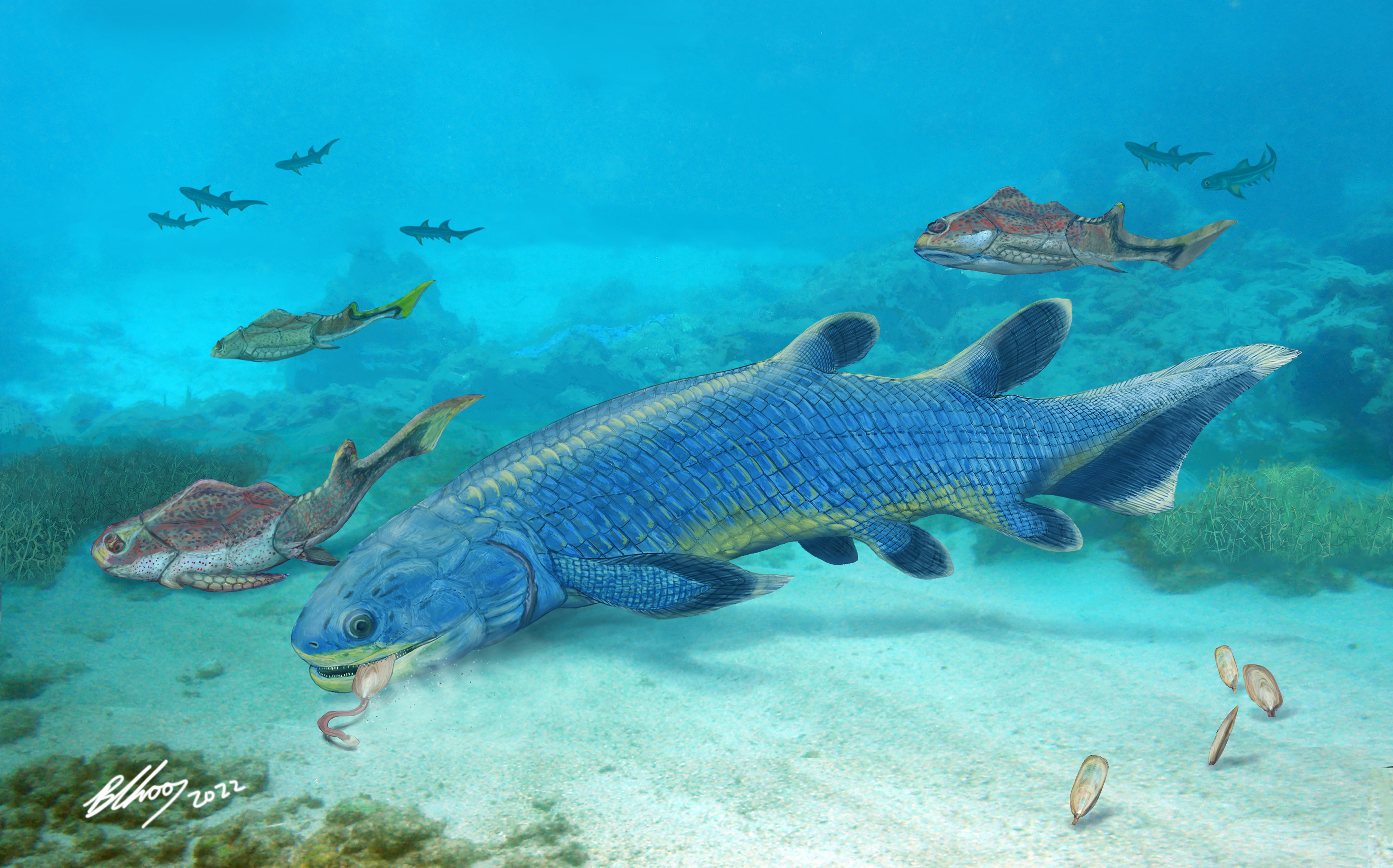
Coelacanths (e.g., Latimeria) are a curious group of sarcopterygian fishes that survive over hundreds of millions of years and are important in evolutionary biology. In the Early Triassic, coelacanths reached their peak of taxonomic diversity but had only patchy fossil record in Asia. Here, we report the discovery of a new species of the coelacanth genus Whiteia on the basis of two specimens from the late Smithian (~ 249 Ma) marine deposits exposed in eastern Anhui, China. The discovery considerably extends the spatial range of Whiteia in the Early Triassic, and documents the oldest species of the genus in Asia, predating the previously oldest record of whiteiids in this continent by nine million years. The new coelacanth with an estimated total length of at least 420 mm, larger than most of other coelacanths (except Rebellatrix) at its age, represents the largest whiteiid named so far from the Early Triassic and provides an important addition for our understanding the evolution of this major Triassic clade of coelacanths.

Paleogeographical distribution of Whiteia in the Early Triassic and reconstructions of head and pectoral girdle in two selected species. 
(a), 1, Whiteia woodwardi, W. tuberculata, and W. uyenoteruyai, Madagascar; 2, W. africana, South Africa; 3, W. gigantea, Texas (USA); 4, Whiteia sp., British Columbia (Canada); 5, W. neilseni, East Greenland; 6, Whiteia anniae sp. nov. Anhui, China.
 (b), reconstruction of head and pectoral girdle of W. woodwardi.

Whiteia anniae sp. nov. in right lateral view, Holotype (CHU 2016).
 (a), Whole specimen. (b), closeup of the calcified lung (indicated by lower arrows) and lateral line (indicated by upper arrows). (c), a scale near the head. (d), anterior tips of jaws with arrows indicating the fangs in anterior coronoids. (e), denticles on the last ray of the anterior dorsal fin.

Systematic palaeontology

Osteichthyes Huxley, 1880.
Sarcopterygii Romer, 1955.
Actinistia Cope, 1871.

Coelacanthiformes Huxley, 1861.

Whiteiidae Schultze, 1993.

Whiteia Moy-Thomas, 193520.

Whiteia anniae Xu, Dai, Tan, Yuan, Sun, Liao, Geng et Song sp. nov.

Etymology. The specific epithet honors the British fossil hunter Mary Anning and her Chinese fan Anni Dai, whose family contributes to the collection of fossils described here.

Holotype. CHU 2016 (Fig. 2). A laterally compressed specimen with the anal fin and caudal missing, stored in the fossil collection of Chaohu University (CHU).
 
Locality and horizon. He County, Anhui Province; Helongshan Formation, Smithian, Olenekian, Early Triassic.

Diagnosis. A large species of Whiteia characterized by the following set of characters (autapomorphies, those unique among Whiteia, identified with an asterisk): presence of six enlarged conical teeth on premaxilla; presence of coronoid fangs (*); presence of contact of first supraorbital with posterior portion of anterior parietal; anterior extremity of preorbital at level of anterior margin of anterior parietal (*); trapezoidal opercle with rounded anteroventral corner; 49 neural arches and 22 haemal arches in vertebral column; eight rays in anterior dorsal fin; pointed denticles associated with rays of anterior dorsal fin; 14 rays and 15 radials, and 12 rays and 13 radials respectively in dorsal and ventral lobes of caudal fin (*); and scale ornamentation consisting of about 20 elongate ridges converging midline posteriorly (*).


Qing-Hua Dai, Guang-Hui Xu, Feng-Ting Tan, Zhi-Wei Yuan, Cheng-Kai Sun, Jun-ling Liao, Bing-He Geng and Hai-Jun Song. 2025. A New coelacanth (Actinistia, Sarcopterygii) from the Early Triassic of Anhui, China. Scientific Reports. 15, 36320. DOI: doi.org/10.1038/s41598-025-20229-w  [17 October 2025]

Friday, November 8, 2024

[PaleoIchthyology • 2024] Graulia branchiodonta • The most detailed anatomical reconstruction of a Mesozoic coelacanth


Graulia branchiodonta
Manuelli, Mondéjar Fernández, Dollman, Jakata & Cavin, 2024


Abstract
Although the split of coelacanths from other sarcopterygians is ancient, around 420 million years ago, the taxic diversity and the morphological disparity of the clade have remained relatively low, with a few exceptions. This supposedly slow evolutionary pace has earned the extant coelacanth Latimeria the nickname “living fossil”. This status generated much interest in both extinct and extant coelacanths leading to the production of numerous anatomical studies. However, detailed descriptions of extinct taxa are made difficult due to the quality of the fossil material which generally prevents fine comparisons with the extant Latimeria. Here we describe a new genus and species of coelacanth, Graulia branchiodonta gen. et sp. nov. from the Middle Triassic of Eastern France, based on microtomographical imaging using synchrotron radiation. Through exquisite 3D preservation of the specimens, we reconstructed the skeletal anatomy of this new species at an unprecedented level of detail for an extinct coelacanth, and barely achieved for the extant Latimeria. In particular, we identified a well-developed trilobed ossified lung whose function is still uncertain. The skeletal anatomy of G. branchiodonta displays the general Bauplan of Mesozoic coelacanths and a phylogenetic analysis resolved it as a basal Mawsoniidae, shedding light on the early diversification of one of the two major lineages of Mesozoic coelacanths. However, despite its exquisite preservation, G. branchiodonta carries a weak phylogenetic signal, highlighting that the sudden radiation of coelacanths in the Early and Middle Triassic makes it currently difficult to detect synapomorphies and resolve phylogenetic interrelationships among coelacanths in the aftermath of the great Permo-Triassic biodiversity crisis.

Graulia branchiodonta gen. et sp. nov.
Photographs of the specimens in laterodorsal view.
(A) MHNG GEPI V5787, holotype. (B) MHNG GEPI V5788.

Systematic paleontology
Class OSTEICHTHYES Huxley, 1880
Subclass SARCOPTERYGII Romer, 1955
Infraclass ACTINISTIA Cope, 1891

Order COELACANTHIFORMES Huxley, 1861
Suborder LATIMERIOIDEI Schultze, 1993

Family MAWSONIIDAE Schultze, 1993

Graulia branchiodonta gen. et sp. nov. 

Holotype: MHNG GEPI V5787, complete specimen

Diagnosis: Mawsoniid coelacanth characterized by the following association of characters: anteromedial process of the posterior parietal present; one pair of lateral extrascapulars; few pores at the sutural contact with bones enclosing the supraorbital sensory canal.; the presence of anterior branches of the supratemporal commissure; a preoperculum with an anterior blade-like portion; a simple anterior end of the lachrymojugal; the infraorbital sensory canal running at the anterior margin of the postorbital; long teeth on the ceratobranchial tooth plates; denticles on the rays of the first dorsal fin and on the first rays of the dorsal lobe of the caudal fin; tri-lobed unpaired lung; scales ornamented with pointed small tubercles. Only known species, same diagnosis as for the genus.

Locality: Sarraltroff, 57400 Moselle, Grand Est, France.

Etymology: The genus name Graulia refers to the Graoully, Graouli or Graully, a mythical dragon from the folklore of Lorraine, the region of France where the specimens were found. The species name branchiodonta, from the greek βράγχια gills” and ὀδούς, ὀδόντος tooth” refers to the large teeth found on the ceratobranchials.

Graulia branchiodonta gen. et sp. nov., interpretative body reconstructions and detailed skull drawings in lateral view.
(A) body profile. (B) lateral view of skull and pectoral girdle. (C) lateral view of skull and pectoral girdle with neurocranium highlighted. (D) lateral view of skull and pectoral girdle with palate and hyobranchial skeleton highlighted.

 
Luigi Manuelli, Jorge Mondéjar Fernández, Kathleen Dollman, Kudakwashe Jakata and Lionel Cavin. 2024.   The most detailed anatomical reconstruction of a Mesozoic coelacanth. PLoS ONE. 19(11): e0312026. DOI: doi.org/10.1371/journal.pone.0312026 

Friday, November 1, 2024

[PaleoIchthyology • 2024] Ngamugawi wirngarri • A Late Devonian coelacanth reconfigures Actinistian Phylogeny, Disparity, and Evolutionary Ddynamics

 
Ngamugawi wirngarri 
Clement, Cloutier, Lee, King, Vanhaesebroucke, Bradshaw, Dutel, Trinajstic & Long, 2024 


Abstract
The living coelacanth Latimeria (Sarcopterygii: Actinistia) is an iconic, so-called ‘living fossil’ within one of the most apparently morphologically conservative vertebrate groups. We describe a new, 3-D preserved coelacanth from the Late Devonian Gogo Formation in Western Australia. We assemble a comprehensive analysis of the group to assess the phylogeny, evolutionary rates, and morphological disparity of all coelacanths. We reveal a major shift in morphological disparity between Devonian and post-Devonian coelacanths. The newly described fossil fish fills a critical transitional stage in coelacanth disparity and evolution. Since the mid-Cretaceous, discrete character changes (representing major morphological innovations) have essentially ceased, while meristic and continuous characters have continued to evolve within coelacanths. Considering a range of putative environmental drivers, tectonic activity best explains variation in the rates of coelacanth evolution.


Ngamugawi wirngarri 
A, B ‘Part a’ of WAM 09.6.148 (holotype) shown in left dorsolateral view and skull close up in left lateral view. C ‘Part b’ of WAM 09.6.148 (holotype) showing all exposed elements; D partial braincase of NMV P231504 (paratype) shown in right lateral view; E cleithrum of NMV P231504 (paratype) in mesial and lateral view; F, G skull reconstruction in dorsal and left lateral view.
Abbreviations: Ang angular, Cl cleithrum, Clv clavicle, Dt dentary, Exc extracleithrum, icj intracranial joint, ioc infraorbital canal, L.Gu lateral gular, Lj lachrymojugal, mc mandibular canal, L.Ex lateral extrascapular, Op operculum, Par Parietal, Po postorbital, Pop preoperculum, Pp postparietal, Pmx premaxilla, Psym parasymphysial, Q quadrate, Ro.p1 anterior pore of the rostral organ, Ro.p2 antero-lateral pore of the rostral organ, Ro.p3 postero-lateral pore of the rostral organ, So supraorbitals, soc supraorbital canal, Sop Suboperculum, Spl splenial, Sq squamosal.

Systematic palaeontology
Osteichthyes Huxley 1880
Sarcopterygii Romer 1955
Actinistia Cope 1871

Ngamugawi wirngarri gen. et sp. nov.

Diagnosis: Ngamugawi wirngarri gen. et sp. nov. is distinguished from all other coelacanths by the following apomorphies: jugal canal with prominent branches; large sensory pore openings between supraorbitals and parietals; teeth on parasymphysial tooth plate, but not on the dentary; prearticular and/or coronoid teeth rounded; cleithra and extracleithra with broad triangular anteroventral overlap for clavicle bearing a large ventral foramen; and scales with long ornamental ridges extending beyond the posterior margin of the base (Figs. 1, 2, Supplementary Fig. 1).

Locality and horizon: Canning Basin, in northern Western Australia, circa 100 km southeast of Fitzroy Crossing; Gogo Formation, early Frasnian, Late Devonian (~384–382 Ma). The holotype was found between Stromatoporoid Camp and Longs Well, the paratype was found in Paddys Valley. 

Etymology: Generic name meaning “ancient fish” in Gooniyandi/Guniyandi, language of the First Nations people from Country around Fitzroy Crossing in the Kimberley region of Western Australia. Specific name is given in honour of respected Gooniyandi elder and ancestor Wirngarri, who lived in the Emanuel Range. Generic and specific names were both provided to Prof. John Long in September 2023, who has a longstanding and ongoing relationship with the community, with permissions to use the language granted by elder Rosemary Nuggett, on behalf of the Gooniyandi people of the Mimbi community.


Phylogenetic relationships and divergence dates within coelacanths, based on tip-dated Bayesian inference.

A live recreation of the Ngamugawi wirngarri coelacanth in its natural habitat. P3D graphic credit: Katrina Kenny. 
 Illustration by Katrina Kenny (courtesy Flinders University)


 Alice M. Clement, Richard Cloutier, Michael S. Y. Lee, Benedict King, Olivia Vanhaesebroucke, Corey J. A. Bradshaw, Hugo Dutel, Kate Trinajstic and John A. Long. 2024. A Late Devonian coelacanth reconfigures Actinistian Phylogeny, Disparity, and Evolutionary Ddynamics. Nature Communications. 15: 7529. DOI: doi.org/10.1038/s41467-024-51238-4


Friday, June 7, 2024

[PaleoIchthyology • 2024] Reconstructing an Ancient Fish: Three-dimensional Skeletal Restoration of the Head of Mawsonia (Sarcopterygii: Actinistia) using CT Scan, and an adjusted model for body size estimation in fossil coelacanths


 Head of Mawsonia (Sarcopterygii, Actinistia)

in Toriño, Dutel, Soto, Norbis, Ezquerra & Perea, 2024. 

Abstract
Mawsonia constitutes one of the most conspicuous fossil coelacanth taxa, due to its unique anatomy and possible maximum body size. It typifies Mesozoic coelacanth morphology, before the putative disappearance of the group in the fossil record. In this work, the three-dimensional cranial anatomy and body size estimations of this genus are re-evaluated from a recently described specimen from Upper Jurassic deposits of Uruguay. The 3D restoration was performed directly on the material based on anatomical information provided by the living coelacanth Latimeria and previous two-dimensional restorations of the head of Mawsonia. The montage was then scanned with computed tomography and virtually adjusted to generate an interactive online resource for future anatomical, taxonomic and biomechanical research. In general terms, the model constitutes a tool to improve both the anatomical knowledge of this genus and its comparison with other coelacanths. It also facilitates the evaluation of possible evolutionary trends and the discussion of particular features with potential palaeobiological implications, such as the anterior position of the eye and the development of the pseudomaxillary fold. Regarding the body size, a previous model for body size estimation based on the gular plate was submitted to OLS, RMA, segmented linear and PGLS regressions (including the evaluation of regression statistics, variance analysis, t-tests and residual analysis). The results point to a power relationship between gular and total lengths showing a better support than a simple linear relationship. The new resulting equations were applied to the studied individual and are provided for future estimates. Although an isometric evolutionary growth cannot be rejected with the available evidence, additional models developed with other bones will be necessary to evaluate possible hidden evolutionary allometric trends in this group of fishes, thus avoiding overestimates.

Keywords: 3D reconstruction, body size, coelacanths, computed tomography, Mawsonia


Pablo Toriño, Hugo Dutel, Matías Soto, Walter Norbis, Víctor Ezquerra and Daniel Perea. 2024. Reconstructing an Ancient Fish: Three-dimensional Skeletal Restoration of the Head of Mawsonia (Sarcopterygii, Actinistia) using CT Scan, and an adjusted model for body size estimation in fossil coelacanths. Journal of Anatomy. DOI: 10.1111/joa.14054 

Thursday, October 26, 2023

[PaleoIchthyology • 2022] Libys callolepis • The First Jurassic Coelacanth from Switzerland


 Libys callolepis 
Ferrante, Menkveld-Gfeller & Cavin, 2022
 

Abstract
Coelacanths form a clade of sarcopterygian fish represented today by a single genus, Latimeria. The fossil record of the group, which dates back to the Early Devonian, is sparse. In Switzerland, only Triassic sites in the east and southeast of the country have yielded fossils of coelacanths. Here, we describe and study the very first coelacanth of the Jurassic period (Toarcian stage) from Switzerland. The unique specimen, represented by a sub-complete individual, possesses morphological characteristics allowing assignment to the genus Libys (e.g., sensory canals opening through a large groove crossed by pillars), a marine coelacanth previously known only in the Late Jurassic of Germany. Morphological characters are different enough from the type species, Libys polypterus, to erect a new species of Libys named Libys callolepis sp. nov. The presence of Libys callolepis sp. nov. in Lower Jurassic beds extends the stratigraphic range of the genus Libys by about 34 million years, but without increasing considerably its geographic distribution. Belonging to the modern family Latimeriidae, the occurrence of Libys callolepis sp. nov. heralds a long period, up to the present day, of coelacanth genera with very long stratigraphic range and reduced morphological disparity, which have earned them the nickname of ‘living fossils’.

Keywords: Sarcopterygii, Actinistia, Libys, New species, Mesozoic, Toarcian, Morphology

Skeleton of Libys callolepis sp. nov. on the part (holotype, NMBE 5034073).
 A Photos with osteological details: 1, denticles on the proximal fin rays of the caudal fin. 2, Postparietal shield with the otic sensory canal opening as a deep groove crossed by pillars (white arrowhead). 3, Posterior parietal and the supraorbitals with their pillars (white arrowhead). 4, Consolidated snout with the anterior opening for the rostral organ (white arrowhead). 5, Teeth on the prearticular. B Semi-interpretative line drawing of the specimen

Libys callolepis sp. nov.

Diagnosis: Libys species with the postparietal shield about half the length of the parietonasal shield (the parietonasal is then proportionally shorter than in the type species). The teeth covering the prearticular are very small, and rounded and smooth. Between 41–47 neural arches. Fin rays are slender than in the type species and then not expanded. The scales are strongly ornamented with irregularly sized and elongated round-to-ovoid ridges disposed along a longitudinal axis.

Etymology: From the ancient Greek καλόςkalós, (‘beautiful’, ‘nice’) and λεπίςlepís, (‘scale’) in reference to the nicely ornamented scales of the species, which differentiates it from the type species.

Holotype and only known specimen: NMBE 5034072 and 5034073, a sub-complete specimen preserved in right lateral view as part and counterpart. Most of the bones, including the scales on the body, are preserved in anatomical position and only the bones of the cheek and the jaw are missing. The specimen is kept in the collections of the Natural History Museum Bern (Canton of Bern, Switzerland).

Horizon and type locality: Toarcian (Lower Jurassic), Creux de l’Ours section, locality of Les Pueys near the Teysachaux summit (Canton of Fribourg, Switzerland).


Skeleton of  Libys callolepis sp. nov. on the counterpart (holotype, NMBE 5034072).
A Photos with osteological details: 1, articular head of the scapulocoracoid. 2, Scales on the flank immediately beneath the first anterior dorsal fin. 3, Scales of the lateral line showing the ornamental pattern with the larger central tubercles (white arrowheads point, showed only on one scale). 4, Scales on the ventral flank from the pelvic to the anal fin. 5, Axial mesomere (white arrowhead) surrounded by some fin rays of the anal fin. 6, Axial mesomeres (white arrowhead) partially covered by sediment in the pelvic fin. B Semi-interpretative line drawing of the specimen


Christophe Ferrante, Ursula Menkveld-Gfeller and Lionel Cavin. 2022. The First Jurassic Coelacanth from Switzerland. Swiss Journal of Palaeontology. 141: 15. DOI: 10.1186/s13358-022-00257-z

Tuesday, October 24, 2023

[PaleoIchthyology • 2023] An enigmatic large mawsoniid coelacanth (Sarcopterygii: Actinistia) from the Upper Jurassic Kimmeridge Clay Formation of England


  Mawsoniidae gen. et sp. indet. (MJML K785).

in Toriño, Gausden, Etches, Rankin, Marshall & Gostling, 2023. 
(artwork by S. Gausden).
 
Abstract
A large mawsoniid coelacanth from the lower part of the marine Kimmeridge Clay Formation of England (Kimmeridgian, Upper Jurassic) is studied here. The material is constituted by a group of bones from the head and shoulder girdle of a considerably large individual (estimated length ca. 1.5 m), including the left angular, left dentary, left prearticular, left palatoquadrate complex, both ceratohyals and right cleithrum. Characters such as the coarse external ornamentation of the angular, and the robustness of the quadrate and the cleithrum allow classification of the individual as a member of the Mawsoniidae; whereas the configuration of external bones of the lower jaw (ornamentation of the angular constituted mainly by longitudinal ridges, the presence of a lateral swelling in the dentary) indicates stronger Gondwanan affinities than previously expected (i.e., with the genus Mawsonia, up to now only recorded in South America by the end of the Jurassic). Considering the above, two alternative evolutionary, paleobiogeographic, and taxonomic scenarios are discussed: (1) the new individual can be referred to the European mawsoniid genus Trachymetopon (Lower–Middle Jurassic), in which case it should be assumed this genus reached the Upper Jurassic, and with a morphological variability higher than previously suspected (including some characters previously assumed as diagnostic for Mawsonia). Or (2) an unknown Mawsonia-like form was present in the Upper Jurassic of Europe. The last scenario puts the identification of isolated elements of European Jurassic giant mawsoniids in a new complex taxonomic and paleobiogeographic context, which will deserve further research.


A, stratigraphic profile of the lower part of the Kimmeridge Clay Formation indicating the level where the material was collected (adapted from Gallois, 2020); B, photograph of the collecting site at Ringstead Bay (one of the authors –S. Etches– for scale).

  Mawsoniidae gen. et sp. indet. (MJML K785).
A, schematic anatomical restoration of the head in left lateral view, showing the preserved bones (missing gray parts based on Maisey 1986; Toriño et al., 2021a); B, hypothetical life restoration
(artwork by S. Gausden).

 A, map showing the geographic provenance of the material studied in this work; B, paleogeographic context during Kimmeridge Clay times, with special reference to Laurasian masses (based on the reconstructions of Cox, 2020 after Callomon, 1985; Thierry, 2000; Gallois, 2020; Stumpf et al., 2021)


Pablo Toriño, Shane F. Gausden, Steve Etches, Kathryn Rankin, John E. A. Marshall and Neil J. Gostling. 2023. An enigmatic large mawsoniid coelacanth (Sarcopterygii, Actinistia) from the Upper Jurassic Kimmeridge Clay Formation of England. Journal of Vertebrate Paleontology. 42(1);  e2125813. DOI: 10.1080/02724634.2022.2125813 

Tuesday, May 3, 2022

[PaleoIchthyology • 2022] The Rapid Evolution of Lungfish Durophagy


Youngolepis praecursor Chang & Yu, 1981
 
in Cui, Friedman, Qiao, et al., 2022. 
artwork by Brian Choo

Abstract
Innovations relating to the consumption of hard prey are implicated in ecological shifts in marine ecosystems as early as the mid-Paleozoic. Lungfishes represent the first and longest-ranging lineage of durophagous vertebrates, but how and when the various feeding specializations of this group arose remain unclear. Two exceptionally preserved fossils of the Early Devonian lobe-finned fish Youngolepis reveal the origin of the specialized lungfish feeding mechanism. Youngolepis has a radically restructured palate, reorienting jaw muscles for optimal force transition, coupled with radiating entopterygoid tooth rows like those of lungfish toothplates. This triturating surface occurs in conjunction with marginal dentition and blunt coronoid fangs, suggesting a role in crushing rather than piercing prey. Bayesian tip-dating analyses incorporating these morphological data indicate that the complete suite of lungfish feeding specializations may have arisen in as little as 7 million years, representing one of the most striking episodes of innovation during the initial evolutionary radiations of bony fishes.

  Youngolepis praecursor, specimen IVPP V28375 in dorsal view.

The palate and dorsal portion of the hyoid arch of Youngolepis praecursor.


Youngolepis praecursor Chang & Yu, 1981

Timing of divergences and rates of trait evolution in lungfishes and their close relatives.


Xindong Cui, Matt Friedman, Tuo Qiao, Yilun Yu and Min Zhu. 2022. The Rapid Evolution of Lungfish Durophagy. Nature Communications. 13: 2390. DOI: 10.1038/s41467-022-30091-3
 highlighting feeding innovations in Youngolepis, an Early Devonian stem lungfish from China (art: Brian Choo) @NatureComms @Friedman_Lab


Friday, November 12, 2021

[PaleoIchthyology • 2021] Mawsonia sp. • The First late Cretaceous Mawsoniid Coelacanth (Sarcopterygii: Actinistia) from North America: Evidence of A Lineage of Extinct ‘Living Fossils’


Reconstruction of Mawsonia sp. roaming in the brackish or fresh water costal environment of Texas during the Cenomanian.

in Cavin, Toriño, Van Vranken, ... et Winkler, 2021. 
Original artwork by Zubin Erik Dutta.

Abstract

Today, the only living genus of coelacanth, Latimeria is represented by two species along the eastern coast of Africa and in Indonesia. This sarcopterygian fish is nicknamed a "living fossil", in particular because of its slow evolution. The large geographical distribution of Latimeria may be a reason for the great resilience to extinction of this lineage, but the lack of fossil records for this genus prevents us from testing this hypothesis. Here we describe isolated bones (right angular, incomplete basisphenoid, fragments of parasphenoid and pterygoid) found in the Cenomanian Woodbine Formation in northeast Texas that are referred to the mawsoniid coelacanth Mawsonia sp. In order to assess the impact of this discovery on the alleged characteristic of "living fossils" in general and of coelacanths in particular: 1) we compared the average time duration of genera of ray-finned fish and coelacanth in the fossil record; 2) we compared the biogeographic signal from Mawsonia with the signal from the rest of the vertebrate assemblage of the Woodbine formation; and 3) we compared these life traits with those of Latimeria. The stratigraphical range of Mawsonia is at least 50 million years. Since Mawsonia was a fresh, brackish water fish with probably a low ability to cross large sea barriers and because most of the continental components of the Woodbine Fm vertebrate assemblage exhibit Laurasian affinities, it is proposed that the Mawsonia’s occurrence in North America is more likely the result of a vicariant event linked to the break-up of Pangea rather than the result of a dispersal from Gondwana. The link between a wide geographic distribution and the resilience to extinction demonstrated here for Mawsonia is a clue that a similar situation existed for Latimeria, which allowed this genus to live for tens of millions of years.


Systematic paleontology
Actinistia Cope, 1871.
Latimerioidei Schultze, 1993.
Mawsoniidae Schultze, 1993.

Genus Mawsonia Woodward in Mawson and Woodward, 1907

Mawsonia sp.


‘Mid’-Cretaceous paleogeographical map of western Laurasia and western Gondwana showing biogeographical affinities of vertebrates from the woodbine formation (red circle).
Brown areas indicates continental vertebrates and the blue areas indicate brackish and marine vertebrates. Orange dots indicate approximate locations of records of Mawsonia.

Comparison of the geographical distributions of the two extant Latimeria species and their supposedly common ancestor 30 to 40 million years ago (left) with those of Mawsonia in the mid-cretaceous, about 100 million years ago and in the Late Jurassic–basal Cretaceous, about 145 million years ago (right).


Lionel Cavin, Pablo Toriño, Nathan Van Vranken, Bradley Carter, Michael J. Polcyn, Dale Winkler. 2021. The First late Cretaceous Mawsoniid Coelacanth (Sarcopterygii: Actinistia) from North America: Evidence of A Lineage of Extinct ‘Living Fossils’ PLoS ONE. 16(11): e0259292. DOI: 10.1371/journal.pone.0259292 


Wednesday, October 25, 2017

[PaleoIchthyology • 2017] Foreyia maxkuhni • Heterochronic Evolution Explains Novel Body Shape in A Triassic Coelacanth from Switzerland


Foreyia maxkuhni 
Cavin, Mennecart, Obrist, Costeur & Furrer, 2017


Abstract

A bizarre latimeriid coelacanth fish from the Middle Triassic of Switzerland shows skeletal features deviating from the uniform anatomy of coelacanths. The new form is closely related to a modern-looking coelacanth found in the same locality and differences between both are attributed to heterochronic evolution. Most of the modified osteological structures in the new coelacanth have their developmental origin in the skull/trunk interface region in the embryo. Change in the expression of developmental patterning genes, specifically the Pax1/9 genes, may explain a rapid evolution at the origin of the new coelacanth. This species broadens the morphological disparity range within the lineage of these ‘living fossils’ and exemplifies a case of rapid heterochronic evolution likely trigged by minor changes in gene expression.


Figure 1: Skeleton of the new coelacanth Foreyia maxkuhni gen. et sp. nov.
(A) Photo and (B) outline of the holotype (PIMUZ A/I 4620). (C) Reconstruction of the whole skeleton.


Sarcopterygii Romer, 1955

Actinistia Cope, 1891

Latimeriidae Berg, 1940 sensu Dutel et al., 20125

Foreyia gen. nov.

Diagnosis: Latimeriid coelacanth with dermal bones covered with numerous large tubercles; hypertrophied otico-occipital portion of skull; fusion of postparietal, supratemporal and extrascapular in postparietal shield, which forms a dome in occipital region; supraorbital sensory canal running in a wide groove; short and curved mandible; pterygopalatine deeper than long with enlarged autopalatine; lachrymojugal and squamosal fused; hypertrophied clavicle; few abdominal vertebrae (seventeen); expanded dorsal and caudal fins; and atrophied pectoral fins.

Foreyia maxkuhni gen. et sp. nov.

Etymology: The generic name honors late Peter L. Forey for his contribution on the study of coelacanth fishes. The specific epithet refers to Max Kuhn, who kindly supported for 12 years the preparation and study of fossils from the Middle Triassic of Graubünden and especially the specimens described here.

Type locality and horizon: Site DF 4 near the Ducanfurgga (Graubünden, Switzerland), upper part of the Prosanto Formation, Middle Triassic (early Ladinian, 240.91 million years ago).


Figure 2: Osteological details of the new coelacanth  Foreyia maxkuhni gen. et sp. nov. (A) Photo and (B) surface CT reconstruction of the skull of the paratype (PIMUZ A/I 4372). (C) Tubercles and denticles in the Holotype (PIMUZ A/I 4620) and (D) in the paratype (PIMUZ A/I 4372).

 1, tubercles on the skull roof. 2, large spine-like tubercles on the posterior margin of the otico-occipital shield. 3, denticles on the fin rays of the first dorsal fin. 4, scales with denticles from the ventral margin of the caudal peduncle. 5, scales with denticles from the anal region. 6, scales with denticles from the belly region. 7, toothed coronoid bones. 8, scales with denticles from the flank. 9, supplementary caudal fin lobe with spiny scales. 10, Scales with denticles from the lobe of the anal fin.

Figure 3: Phylogenetic relationships of  Foreyia maxkuhni gen. et sp. nov. and developmental origin of the derived characters.
(A) Strict consensus trees of the 259 most parsimonious trees of 317 steps (CI = 0.3817, RI = 0.6766) with some of the uniquely derived characters present in Foreyia maxkuhni on the left, and reconstructions of genera with atypical general morphology. (B and C) Shared features of Ticinepomis peyeri and Foreyia maxkuhni (in orange) not included in the cladistics analysis (see main text for numbers). (D) Reconstruction of a coelacanth embryo with localization of embryonic tissues that give rise the derived skeletal features present in Foreyia. It is hypothesized that changes in the expression of Pax9 may have altered the derived characters shown in blue on the reconstruction (E).
All the drawings were made by LC.  Abbreviation: Boc, basioccipital; Cla, clavicle; Exo, exoccipital; lat. Meso., lateral mesoderm; neur. cr., neural crest; pect. f., pectoral fin; S (numbered), somite.

 Reconstruction of the living coelacanth  Foreyia maxkuhni gen. et sp. nov.
 Artwork by Alain Bénéteau.

Lionel Cavin, Bastien Mennecart, Christian Obrist, Loïc Costeur and Heinz Furrer. 2017. Heterochronic Evolution Explains Novel Body Shape in A Triassic Coelacanth from Switzerland.  Scientific Reports. 7,  13695.  DOI:  10.1038/s41598-017-13796-0