Showing posts with label Journal: Acta Palaeontol. Pol.. Show all posts

Saturday, July 11, 2026

[PaleoIchthyology • 2026] The Oldest Shark Face—Anatomy of the Devonian elasmobranch Phoebodus


Phoebodus saidselachus Frey, Coates, Ginter, Hairapetian, Rücklin, Jerjen & Klug, 2019

in Klug, Greif, Pohle, Ginter, Coates, Haouz, Lagnaoui, Pople et Frey, 2026. 

Phoebodus was recognized as the earliest elasmobranch known from articulated remains, a group which constitutes most modern cartilaginous fish comprising sharks, skates and rays. Its elongate body, the presence of two dorsal fins with fin spines, and the elongate head had already been described. Based on spectacularly well-preserved fossils, we add new anatomical information on its exact body proportions, the paired and caudal fins, the dermal denticles, the skull morphology, the endocast, and the gill skeleton. These new skeletons from the Famennian (Upper Devonian) of Morocco (c. 367 Ma) permit a much-improved reconstruction of the anatomy of Phoebodus. The new materials comprise the oldest elasmobranch specimens preserving the complete head in three dimensions. Additionally, the new materials yield information about growth and diet and thus position in the trophic network. Despite the newly coded characters, the phylogenetic position of the genus Phoebodus with the oldest teeth dating to the early Givetian remains that of the oldest stem-elasmobranch in the Bayesian analyses.

Key words: Chondrichthyes, Elasmobranchii, Famennian, ontogeny, exceptional preservation, Fossillagerstätten

Phoebodontiform elasmobranch Phoebodus saidselachus Frey et al., 2019a, middle Famennian, Upper Devonian, southern Maïder, Anti-Atlas, Morocco. A. PIMUZ A/I 5751, prep. T. Imhof, largest individual, nearly complete. B. PIMUZ A/I 5752, prep. T. Imhof, second largest individual, only caudal region missing; for details of the skull see Fig. 2. C. PIMUZ A/I 5753, prep. M. Greif, anterior half, excellent integument, fins and brachial region. D. PIMUZ A/I 5754, prep. R. Roth, two superimposed chondrichthyans; the skull in the middle is Maghriboselache mohamezanei with further remains; the straight, complete skeleton belongs to Phoebodus; the latter preserves the caudal fin. E. PIMUZ A/I 4712, holotype, smallest skeleton, nearly complete, modified after Frey et al. (2019a).

Skeletal reconstruction of the phoebodontiform elasmobranch Phoebodus saidselachus Frey et al., 2019a, middle Famennian, Upper Devonian, southern Maïder, Anti-Atlas, Morocco. A. PIMUZ A/I 5754, caudalis. B, C. PIMUZ A/I 4712, posterior and anterior dorsal fins. D. PIMUZ A/I 5752, anterior dorsal fin. E. PIMUZ A/I 5751, caudalis. F, G. PIMUZ A/I 5753, pectoral fin and branchial basket. H. PIMUZ A/I 5752, head. The photos are not to scale. The scale refers only to the drawing.

Systematic palaeontology 
Chondrichthyes Huxley, 1880 
Elasmobranchii Bonaparte, 1838 
Phoebodontiformes Ginter et al., 2010 

Genus Phoebodus St. John & Worthen, 1875


Reconstructions of an adult female and juvenile male of the phoebodontiform elasmobranch Phoebodus saidselachus Frey et al., 2019a, middle Famennian, Upper Devonian, southern Maïder, Anti-Atlas, Morocco. We incorporated the new anatomical details particularly in the proportions and the fin shapes. The juvenile is reconstructed after the holotype.

  
Christian Klug, Merle Greif, Alexander Pohle, Michał Ginter, Michael I. Coates, Wahiba Bel Haouz, Abdelouahed Lagnaoui, Jonathan Pople, and Linda Frey. 2026. The Oldest Shark Face—Anatomy of the Devonian elasmobranch Phoebodus. Acta Palaeontologica Polonica. 71 (2), 2026: 399-430 DOI:10.4202/app.01290.2025  https://www.app.pan.pl/article/item/app012902025.html

Linda Frey, Michael Coates, Michał Ginter, Vachik Hairapetian, Martin Rücklin, Iwan Jerjen, Christian Klug. 2019. The early elasmobranch Phoebodus: phylogenetic relationships, ecomorphology and a new time-scale for shark evolution. Proc Biol Sci. 286 (1912): 20191336. DOI: doi.org/10.1098/rspb.2019.1336

Thursday, June 4, 2026

[Paleontology • 2026] Patagoniaemys aeschyli • A New meiolaniform Turtle from the Maastrichtian of Northern Patagonia, Argentina


Patagoniaemys aeschyli
Agnolin, Rolando, Sterli, Chimento, Novas & Muñoz, 2026


Meiolaniformes are a group of chelonians including the famous horned-turtles Niolamia argentina (Patagonia) and Meiolania platyceps (Australia). In South America, the Late Cretaceous meiolaniforms are represented by two named taxa: Patagoniaemys gasparinae coming from Campanian–Maastrichtian beds of the La Colonia Formation, Chubut province, and Trapalcochelys sulcata from the Campanian–Maastrichtian beds of the Allen Formation, Río Negro Province. The aim of the present contribution is to describe a new meiolaniform chelonian coming from the Upper Cretaceous (Maastrichtian) Los Alamitos Formation, at Río Negro Province, Argentina. The material is represented by a partial basicranium, incomplete carapace and fragmentary postcranial bones (MPMIK 1839/P/33) belonging to a new species Patagoniaemys aeschyli. To this new species we refer all the material previously referred as indeterminate meiolaniid, meiolaniform and cf. Niolamia sp., coming from the same site and locality, and described by different authors since the 1980s. This new species differs from the type species P. gasparinae, particularly by the presence of longitudinal ridges in the nuchal bone. We also include brief comments about meiolaniform palaeobiogeography and the impact of K/Pg extinction event in Patagonian chelonians.

Key words: Testudinata, Meiolaniformes, Los Alamitos Formation, Maastrichtian, Cretaceous, Patagonia, Argentina.


Meiolaniform turtle Patagoniaemys aeschyli sp. nov. holotype (MPMIK 1839/P/33); Cerro Cuadrado locality, Argentina, Los Alamitos Formation Maastrichtian (Upper Cretaceous).
A. Carapace; anterior border in dorsal (A1, A2) and ventral (A3, A4) views, detail of the anterior margin in ventral view (A7, A8), showing the presence of longitudinal ridges that characterize the new species (light grey, nuchal bone; dark grey, broken surfaces).
B. Left hyoplastron fragment Scale bars 100 mm.

Testudinata Klein, 1760 
Meiolaniformes Sterli & de la Fuente, 2013 

Genus Patagoniaemys Sterli & de la Fuente, 2011a 
Type species: Patagoniaemys gasparinae Sterli & de la Fuente, 2011a;
Chubut Province, Argentina, Campanian–Maastrichtian. 

Patagoniaemys aeschyli sp. nov.

Diagnosis.—Large chelonian (maximum anterior carapace width about 60 cm, maximum inferred carapace length 80 cm). Referable to meiolaniforms by several features, namely the presence of small pits and grooves ornamenting carapace bones, anteriorly curved grooves among marginal scutes, opisthocoelous caudal vertebrae, and carapace-plastron connection through ligaments (Gaffney 1996; Hirayama et al. 2000; Sterli and de la Fuente 2013; Sterli et al. 2013). It is referred to Patagoniaemys by having a basioccipital with well-developed basal tubera with a concave surface between them, posteriorly flat occipital condyle, a shallowly notched anterior carapace margin, presence of a thickened bump on ...

Etymology: In reference to Aeschylus (525–455 BC), an ancient Greek writer often described as the father of tragedy. During a visit to the city of Gela (Italy), he was killed outside the city by a tortoise dropped by an eagle, which had mistaken his bald head for a rock suitable for shattering the shell.


the excavation work at the Los Alamitos Formation southwest of Cerro Cuadrado.  


Federico L. Agnolin, Mauro Aranciaga Rolando, Juliana Sterli, Nicolás R. Chimento, Fernando E. Novas, and Gonzalo L. Muñoz. 2026. A New meiolaniform Turtle from the Maastrichtian of Northern Patagonia, Argentina. Acta Palaeontologica Polonica. 71(1); 173-184. DOI: doi.org/10.4202/app.01268.2025 

DESCUBREN UNA TORTUGA GIGANTE DEL FIN DE LA ERA DE LOS DINOSAURIOS EN PATAGONIA

Monday, March 23, 2026

[Paleontology • 2026] Balticolasma wunderlichi • 3D analyses of the First ortholasmatine Harvestmen (Opiliones: Nemastomatidae) from European Eocene Ambers


Balticolasma wunderlichi 
Bartel, Mitov, Dunlop & Hammel, 2026

Artistic reconstruction by Joshua Knüppe  x.com/JoschuaKnuppe


The first fossil representatives of the harvestman subfamily Ortholasmatinae (Opiliones, Dyspnoi, Nemastomatidae) are described as Balticolasma wunderlichi gen. et sp. nov. One male is preserved in Eocene Baltic amber and a presumably conspecific female in Eocene Rovno amber (northwest Ukraine). Ortholasmatines are typically highly ornate arachnids, and for the first time with an amber harvestman we applied computed tomography using synchrotron radiation to investigate its three-dimensional morphology and surface structure in considerable detail. Some of its morphological characters, appear to be closer to extant Asian genera. In a wider biogeographic context, our amber record is a significant find for the Paleogene of Europe given that (i) it is another species apparently found in both Baltic and Rovno amber and (ii) all modern ortholasmatines are restricted to East Asia and North and Central America.

Key words: Nemastomatidae, Ortholasmatinae, Baltic amber, Rovno amber, micro-CT, Priabonian, Eocene

Ortholasmatine harvestman Balticolasma wunderlichi gen. et sp. nov. paratype (female) MB.A.4455 from Rovno amber, 33.90–37.71 Ma, Priabonian.
A1, dorsal view of the body A2, close-up of the ocular process in dorsal view, eye lenses arrowed.
Abbreviations: cp, carapace process; ey, eye lens; oc, ocular process; legs numbered I–IV. Scale bars 1 mm (A1) and 0.5 mm (A2).

Artistic reconstruction of the Eocene ortholasmatine harvestman Balticolasma wunderlichi gen. et sp. nov. (male) by Joshua Knüppe (Ibbenbüren, Germany).

Systematic palaeontology 
Order Opiliones Sundevall, 1833 
Suborder Dyspnoi Hansen & Sørensen, 1904 
Family Nemastomatidae Simon, 1872 
Subfamily Ortholasmatinae Shear & Gruber, 1983 
Genus Balticolasma nov.

Balticolasma wunderlichi gen. et sp. nov. 

Etymology: From the Baltic region where the amber hosting one of the specimens originates from and the suffix lasma applied to a few possibly closely related modern ortholasmatine genera.


Christian Bartel, Plamen G. Mitov, Jason A. Dunlop, and Jörg U. Hammel. 2026. 3D analyses of the First ortholasmatine Harvestmen from European Eocene Ambers. Acta Palaeontologica Polonica. 71(1): 95-107. DOI: doi.org/10.4202/app.01283.2025

Wednesday, December 24, 2025

[Paleontology • 2025] Mandocaris polyphaga • A New caridean Shrimp Fossil (Caridea: Acanthephyridae) with exceptionally preserved organs from the Middle Jurassic of La Voulte-sur-Rhône, France


Mandocaris polyphaga
Lagrange, Audo, Odin, De Grave, Fernandez, Dollman & Charbonnier, 2025


We used propagation phase contrast synchrotron X-ray micro-computed tomography (PPC-SRμCT) on an exceptionally preserved fossil caridean from the Callovian of the La Voulte-sur-Rhône Konservat-Lagerstätte. The tomographic data reveal the shape of the mandible and pereiopodal epipods allowing the description of a new genus and species of Acanthephyridae (Caridea) shrimp, Mandocaris polyphaga gen. et sp. nov. Most organs are exceptionally preserved in either mineral denser to X-ray than matrix, interpreted to be sulfides, or in mineral of lower density than the matrix, interpreted as carbonate/phosphate such as fluorapatite. We herein propose a taphonomic scenario for the preservation of M. polyphaga gen. et sp. nov.: it died from unknown causes not caused by an injury, as no wound is visible, falling on the sediment/water interface, it laid on its right side, and was probably covered by sediments and/or a microbial mat, thus quickly becoming entombed in the anoxic zone of the sedimentary column. Once there, many anatomic structures were replaced by phosphates. Sulfides precipitated concomitantly or quickly afterwards, probably aided by both internal and external source of metal ions. The importance of the external source of metal ions (hydrothermalism) is clear due to the prevalence of sulfides in the ventral side of the specimen, an area more permeable due to its abundance in thin membranes prone to decay. The loss of integrity thereafter led to sediment invading the body cavity, thus obliterating a few ventral anatomic details, including some pereiopodal muscles, part of the hepatopancreas, most of the gills, and possibly reproductive organs. The nodule was then formed, closing the system, and protecting the specimen from further diagenetic degradation.

Key words: Crustacea, Caridea, synchrotron, tomography, anatomy, Konservat-Lagerstätte, Callovian, Middle Jurassic.

Overview of the caridean shrimp Mandocaris polyphaga gen. et sp. nov. (holotype, MNHN.F.A58277) from the Callovian (Middle Jurassic) of La Voulte, France.
Specimen in right lateral (A1) and dorsal (A2) views. Cephalothorax in right lateral view (A3). 3D reconstruction of the holotype in right lateral view (A4), exposing the organs (A5). Abbreviations: a, branchiocardiac groove; a1, antennula; a2, antenna; b1, hepatic groove; e1e, cervical groove; hc, hepatic carina; hs, hepatic spine; mxp3, third maxilliped; phc, posterior hepatic carina; P1–5, pereiopods 1–5; pl1–4, pleopods 1–4; s1–6, pleonites 1–6. White arrows point to the front of the animal.

Mandocaris polyphaga gen. et sp. nov. 
 

Flavien Lagrange, Denis Audo, Giliane P. Odin, Sammy De Grave, Vincent Fernandez, Kathleen Dollman, and Sylvain Charbonnier. 2025. A New caridean Shrimp Fossil with exceptionally preserved organs from the Middle Jurassic of La Voulte-sur-Rhône, France. Acta Palaeontologica Polonica. 70(4); 775-794. DOI: 10.4202/app.01275.2025

Thursday, December 4, 2025

[Paleontology • 2025] Falciscaris mumakiana • A New giant nektobenthic radiodont benthivore (Radiodonta: Hurdiidae) from the Early Ordovician Fezouata Biota in Morocco


Falciscaris mumakiana 
Potin, Claisse, Trébaol, Gueriau, Wu, Pates & Daley, 2025

Artwork by Alexis Trébaol

The Fezouata Shale Formation is an Early Ordovician Lagerstätte that preserved exceptionally detailed records of complex marine ecosystems, making it crucial for understanding the early evolution of animal life. It has yielded the youngest known community of radiodonts to date. This group is particularly well known from the Cambrian, with iconic representatives such as Anomalocaris, which are emblematic of the Cambrian explosion. Here we describe a new radiodont from the Fezouata Biota, Falciscaris mumakiana gen. et sp. nov. based on seven specimens of isolated frontal appendages. These appendages bear long endites with large and robust auxiliary spines, suggesting they were adapted for foraging through sediment in search of prey. The appendages of F. mumakiana gen. et sp. nov. can be relatively large compared to the majority of radiodont appendages, with endites reaching up to 11.4 cm in length, suggesting a total body size exceeding one meter for this Ordovician radiodont. In contrast, smaller specimens can be up to 10 times smaller, indicating ontogenetic stages during which the frontal appendage morphology changes little. Following the “Ordovician Plankton Revolution”, the proliferation of planktonic resources and enhanced pelagic-benthic coupling during this period likely allowed for the rise of giant suspension-feeding radiodonts, such as the Aegirocassisinae and F. mumakiana gen. et sp. nov., the new giant benthivore. In term of taxonomic diversity, benthivores radiodonts remain a minor component of radiodont diversity in the Fezouata Biota compared to the more dominant suspension feeders.

Key words: Panarthropoda, Radiodonta, Hurdiidae, gigantism, benthivores, feeding evolution, Fezouata Shale, Early Ordovician.


Superphylum Panarthropoda Nielsen, 1995 
Order Radiodonta Collins, 1996 
Family Hurdiidae Lerosey-Aubril & Pates, 2018

Hurdiid radiodont Falciscaris mumakiana gen. et sp. nov. frontal appendage, 
from the Fezouata Shale Formation, Lower Ordovician, Morocco.
YPM IP 516782a/b, holotype and paratypes 

 Genus Falciscaris nov.

Etymology: From Latin falx (genitive falcis), scythe, in recognition of the strongly curved shape of the endites tips; and Latinised Greek cariscrab, commonly used in arthropod taxonomy.

Falciscaris mumakiana sp. nov.  

 Etymology: Mûmak (plural mûmakil) is a fantasy elephant-like animal from the Tolkien universe Lord of the Rings. They are described as giant animals and are depicted in the third part of the movie trilogy as having 4 long curved tusks equipped with spines, looking like the curved and spiny endite of Falciscaris mumakiana gen. et sp. nov. 

Diagnosis.—Hurdiidae frontal appendage with at least seven podomeres: one proximal, five intermediate and one distal. Proximal podomeres rectangular, taller than long. At least six laminiform endites are present, long ones on five intermediate podomeres and a shorter one distally. Intermediate endites over twice the height of podomeres, all ending in a strongly curved, hook-like tip. Each endite bears dorsally curved auxiliary spines in at least three sizes, alternating such that spines of the same size are never adjacent.

Artistic life reconstruction of the hurdiid radiodont Falciscaris mumakiana gen. et sp. nov. 
Apart from the frontal appendages, the reconstruction is based on other related hurdiids.
by Alexis Trébaol


Gaëtan J.-M. Potin, Pénélope Claisse, Alexis Trébaol, Pierre Gueriau, Yu Wu, Stephen Pates, and Allison C. Daley. 2025. A New giant nektobenthic radiodont benthivore from the Early Ordovician Fezouata Biota in Morocco. Acta Palaeontologica Polonica. 70(4). 709-722. DOI: doi.org/10.4202/app.01278.2025 

Monday, November 10, 2025

[Paleontology • 2025] Gadusaurus aqualigneus • A New ichthyosaur from the Lower Jurassic of Portugal (Iberian Peninsula)


Gadusaurus aqualigneus 
 Sousa, Roldão, Ríos & Puértolas-Pascual, 2025
    

Ichthyosaurs were a group of marine Mesozoic reptiles. Their presence in the Portuguese fossil record is scarce and ranges from the Sinemurian to the Toarcian. In this work we describe a new specimen—a nearly complete skull discovered in the Sinemurian of Praia de Água de Madeiros, São Pedro de Moel, Municipality of Marinha Grande (Portugal). This specimen represents the most complete and best preserved cranial remains from the Iberian Peninsula published to date. Phylogenetic analyses performed identified this specimen as a new genus and species, Gadusarus aqualigneus, the first to be identified in the Iberian Peninsula. The species is phylogenetically recovered as a baracromian ichthyosaur, part of the Neoichthyosauria and Parvipelvia clades.

Key words: Parvipelvia, Ichthyosauria, Gadusaurus aqualigneus , phylogeny, Sinemurian, São Pedro de Moel.

 Ichthyosaur reptile Gadusaurus aqualigneus gen. et sp. nov. (ML 2750) from from the Lower Jurassic of São Pedro de Moel (Marinha Grande, Portugal). Nearly complete skull in laterodorsal view. 

Ichthyopterygia Owen, 1859 
Ichthyosauria Blainville, 1835 
Parvipelvia Motani, 1999 
Neoichthyosauria Sander, 2000 
Thunnosauria Motani, 1999 
Baracromia Fischer et al., 2013

 Genus Gadusaurus nov.  
Type species: Gadusaurus aqualigneus sp. nov. 

Etymology: From combination of Latin gaducodfish, and Ancient Greek, sauroslizard; in reference to both the ichthyosaur fish-like body shape, and the proclivity for codfish in Portuguese cuisine. 
 
Gadusaurus aqualigneus sp. nov.  

 Etymology: From Latin aquawater and lignumwood; in reference to Água de Madeiros, which roughly translates to “water of woods”, the beach where the specimen was found. 

Holotype: ML 2750, nearly complete skull (Fig. 2), in a flattened state, the right side fully observable in lateral view, and elements of the left side observable in dorsal view. 

Type locality: Praia de Água de Madeiros, São Pedro de Moel, Municipality of Marinha Grande, (Portugal). 

Type horizon: Upper part of the Polvoeira Member of the Água de Madeiros Formation, upper Sinemurian (Lower Jurassic). 

Diagnosis.—Small-sized ichthyosaur, with an estimated length of approximately 2 m, diagnosed by the following combination of ambiguous synapomorphies: absence of a supranarial process in the premaxilla; and the parietal foramen being entirely surrounded by the frontals. Gadusaurus aqualigneus gen. et sp. nov. is also characterized by the following potential autapomorphies within Parvipelvia: presence of a subcircular depression in the lacrimal just below the posterior region of the external naris (Fig. 3C); big and well-developed elliptical internasal foramen with about 5 times bigger surface area than that of the parietal foramen (Fig. 3A); presence of a well-marked longitudinal groove on each side of the anterolateral region of the excavatio internasalis (Fig 3B).


João Pratas E Sousa, Isabel Morais Roldão, María Ríos, and Eduardo Puértolas-Pascual. 2025. A New ichthyosaur from the Lower Jurassic of Portugal (Iberian Peninsula). Acta Palaeontologica Polonica. 70(1); 179-192. DOI: doi.org/10.4202/app.01199.2024

Wednesday, April 9, 2025

[PaleoMammalogy • 2025] Ravjaa ishiii • New Late Cretaceous zhelestid mammal (Eutheria: Zhelestidae) from the Bayanshiree Formation, Mongolia


Ravjaa ishiii 
Okoshi, Takasaki, Chiba, Natori, Saneyoshi, Takahashi, Kodaira, Hayashi, Ishigaki, Mainbayar & Tsogtbaatar, 2025


Numerous exquisitely preserved mammal fossils unearthed from Upper Cretaceous strata in the Gobi Desert of Mongolia have played a key role in understanding Mesozoic mammalian evolution. These splendid mammal fossils have been recovered mainly from the Baruungoyot and Djadokhta formations, but only two fragmentary remains of mammals have been collected from the underlying Bayanshiree Formation. Here we report a new species of Zhelestidae, Ravjaa ishiii gen. et sp. nov., based on a new mammal specimen recently discovered from the Bayanshiree Formation at the Bayan Shiree locality. The new specimen is represented by a well-preserved partial right dentary with the distal portion of an ultimate premolar and the first to the third molars. The gross observation and the phylogenetic analyses demonstrate zhelestid affinities: the subequally tall protoconid and metaconid, closely approximated hypoconulid and entoconid. Zhelestidae was widely distributed from Eurasia to North America in the Late Cretaceous but has not previously been recovered in the abundant Late Cretaceous mammalian fauna of Mongolia. This species represents the first zhelestid from the Mongolian Upper Cretaceous strata and contributes to further understanding of their paleobiogeographic and ecological insights.

Key words: Mammalia, Eutheria, Cretaceous, Bayanshiree Formation, Mongolia.


. Holotype of the zhelestid eutherian mammal Ravjaa ishiii gen. et sp. nov. (MPC-M 100s/001) from the Upper Cretaceous Bayanshiree Formation, Dornogovi, Mongolia.
Right mandible in labial (A), occlusal (B) and lingual (C) views.
Photographs (A1–C1) and 3D model (A2–C2) produced from a CT scan.
 
Mammalia Linnaeus, 1758 
Theria Parker and Haswell, 1897 
Eutheria Gill, 1872 
Zhelestidae Nessov, 1985a 

Genus Ravjaa nov. 

 Etymology: Derived from the 5th Noyon Khutugt lama of the Gobi, Dulduityn Danzanravjaa, the famous Buddhist scholar who was deeply respected by people in the Dornogobi region, where this specimen was unearthed. Given that the genus name “Ravjaa” originates from the personal name of a male figure, it should be considered masculine. 

Ravjaa ishiii sp. nov. 

Etymology: In honor of Ken-ichi Ishii, the director of Hayashibara Museum of Natural Sciences, Okayama, Japan, for his leading role in the Japanese team of the Mongolia-Japan joint expedition, which tremendously contributes to Mongolian paleontology. Holotype: MPC-M 100s/001, partial right dentary with a distal portion of the ultimate premolar, damaged m1, and well-preserved m2–3. 

Type locality: Bayan Shiree, located approximately 25 km southwest of Dzüünbayan, Dornogovi, Mongolia. 

Type horizon: Cenomanian–Santonian, Upper Cretaceous, Bayanshiree Formation. 

Diagnosis.—A zhelestid eutherian that differs from all other zhelestids in: the faint coronoid crest of the dentary with the shallow masseteric fossa, the concave dorsal alveolar margin of the dentary, the complete absence of the labial mandibular foramen, and the significantly high m2 crown that reaches approximately 70% of the height of the horizontal ramus directly below it on labial view.


Tsukasa Okoshi, Ryuji Takasaki, Kentaro Chiba, Masahito Natori, Mototaka Saneyoshi, Akio Takahashi, Shota Kodaira, Shoji Hayashi, Shinobu Ishigaki, Buuvei Mainbayar, and Khishigjav Tsogtbaatar. 2025. New Late Cretaceous zhelestid mammal from the Bayanshiree Formation, Mongolia. Acta Palaeontologica Polonica. 70 (1); 193-203. DOI: doi.org/10.4202/app.01213.2024

Tuesday, June 4, 2024

[Paleontology • 2024] Musankwa sanyatiensis • A new Late Triassic sauropodomorph dinosaur (Sauropodomorpha: Massopoda) from the Mid-Zambezi Basin, Zimbabwe


Musankwa sanyatiensis
 Barrett, Chapelle, Sciscio, Broderick, Zondo, Munyikwa & Choiniere, 2024


An articulated partial hind limb collected from the Pebbly Arkose Formation (Norian, Upper Triassic) of the Upper Karoo Group of Zimbabwe is described as a new taxon of sauropodomorph dinosaur. Musankwa sanyatiensis gen. et sp. nov. was discovered on the shoreline of Lake Kariba, on Spurwing Island in the Mid-Zambezi Basin. The holotype consists of a right femur, tibia, and astragalus, and can be distinguished from all other Late Triassic massopodan sauropodomorphs on the basis of numerous features, which form a unique character combination. Phylogenetic analysis recovers the new taxon as the earliest-branching lineage within Massopoda. Musankwa is only the fourth dinosaur to be named from the Karoo-aged basins of Zimbabwe and further demonstrates the high potential of this region for discoveries of new early dinosaur material.

Key words: Sauropodomorpha, Massopoda, Pebbly Arkose Formation, Norian, Lake Kariba.


Sauropodomorpha Huene, 1932 
Massopoda Yates, 2007 

Genus Musankwa nov.

Etymology: In reference to the house-boat “Musankwa”, from the Tonga dialect, meaning “boy close to marriage”; see SOM 5: fig. S1. This vessel acted as our home and mobile laboratory during two field expeditions to Lake Kariba in 2017–2018, and was made available through the generosity of David and Julie Glynn. Its crew, Coster Katupu, Godfrey Swalika, Simbarashe Mangoroma, and Never Mapira, provided essential logistic support.


Musankwa sanyatiensis gen. et sp. nov.

Etymology: Named for the Sanyati River, whose original course is now submerged in the Sanyati Basin of Lake Kariba, in which the type locality (Spurwing Island) is situated. 


Paul M. Barrett, Kimberley E. J. Chapelle, Lara Sciscio, Timothy J. Broderick, Michel Zondo, Darlington Munyikwa, and Jonah N. Choiniere. 2024. A new Late Triassic sauropodomorph dinosaur from the Mid-Zambezi Basin, Zimbabwe. Acta Palaeontologica Polonica 69 (2); 227-241. DOI: 10.4202/app.01100.2023

Friday, April 5, 2024

[Paleontology • 2024] Pterosaur Teeth from the Southern Neuquén Basin (Patagonia, Argentina): New Insights on the Reconstruction of Ornithocheiriform Dental Anatomy


 Life reconstruction of the Cerro de los Leones pterosaur, displaying the morphological diversity of its dentition based on the observed distribution of the morphotypes.

in Ciaffi et Bellardini, 2024. 
Illustration by Alessio Ciaffi.   twitter.com/CiaffiAlessio

The pterosaur fossil record of Argentina is increasing in recent times, both in the number of localities and their temporal range. The new materials are found in levels that span from the Late Triassic to the Late Cretaceous in age. In this contribution we describe twenty isolated pterosaur teeth from the Cerro de los Leones locality (Picún Leufú, Neuquén Province), where the Lohan Cura Formation (Albian) widely crops out. A new, detailed analysis of these remains allowed us to detect a set of morphological features, such as the enamel texture, cross-section shape, crown curvature, and absence of sharp carinae, which suggests close affinities with Ornithocheiriformes. Moreover, with the large number of tooth specimens, a discrimination of three morphotypes based on the crown curvature, the basal cross-section shape, and the presence of carinae was carried out. We performed extensive morphological comparisons between different members of Ornithocheiriformes with the aim to test the hypothesis that the presence of these different morphotypes reflects distinct anatomical positions of the teeth in the jaws . Thus, considering the heterodont dentition of ornithocheiriforms, the tooth morphological classification proposed here represents a new methodology, not only to identify isolated ornithocheiriform teeth, but also to assign them to the rostral, medial, or the posterior portion of the tooth row. Finally, the evidence of ornithocheiriform pterosaurs in Cerro de los Leones has paleobiogeographical implications, allowing us to correlate it with other Albian faunas from South America, expanding our knowledge of the distribution of Early Cretaceous pterosaurs across southwestern Gondwana.

Key words: Anhangueridae, Cretaceous, Gondwana, Neuquén Basin, tooth morphology, enamel, heterodonty.

Systematic paleontology 
Pterosauria Kaup, 1834 
Pterodactyloidea Plieninger, 1901 
Lanceodontia Andres et al., 2014 
Ornithocheiriformes Andres, 2021


Alessio Ciaffi and Flavio Bellardini. 2024. Pterosaur Teeth from the Southern Neuquén Basin (Patagonia, Argentina): New Insights on the Reconstruction of Ornithocheiriform Dental Anatomy. Acta Palaeontologica Polonica. 69 (1): 73-86. DOI: 10.4202/app.01122.2023
 

Wednesday, June 14, 2023

[Paleontology • 2023] Wulong bohaiensis • Iridescent Plumage in A juvenile dromaeosaurid Theropod Dinosaur


Wulong bohaiensis  Poust, Gao, Varricchio, Wu & Zhang, 2020

in Croudace, Shen, Lü, Brusatte eVinther, 2023. 
Artwork by Robert Nicholls (Bob Nicholls Art).

Colour reconstructions have provided new insights into the lives of dinosaurs and other extinct animals, by predicting colouration patterns from fossilised pigment-bearing organelles called melanosomes. Although these methods have become increasingly popular, only a small number of dinosaurs have been studied using these techniques, which require exceptional preservation of fossil feathers, leaving open key questions such as whether dinosaurs changed their plumage patterns during ontogeny. Here we reconstruct the feather colouration of an approximately one-year-old individual of the Early Cretaceous dromaeosaurid theropod Wulong bohaiensis, which to our knowledge is the first unequivocal juvenile paravian for which aspects of the original colour has been predicted. Using quadratic discriminant analysis (QDA) and multinomial logistic regression (MLR) on the most comprehensive available datasets, we find strong evidence for iridescent plumage of the forelimb and hindlimb remiges and grey plumage on other portions of the body. This suggests that some juvenile paravians used shiny iridescent feathers for signalling purposes, possibly even before reaching somatic or sexual maturity, and thus we can conclude that this paravian used iridescent signalling for intraspecific communication other than sexual signalling. Finally, our results show that when analysing fossil datasets that are entirely comprised of solid and cylindrical melanosomes QDA consistently outperforms MLR, providing more accurate and higher classification probability colour predictions.

Key words: Dinosauria, Dromaeosauridae, Paraves, Wulong bohaiensis , iridescence, melanosome, palaeocolour, juvenile, colour reconstruction, Cretaceous, China.

Reconstruction of the dromaeosaurid dinosaur Wulong bohaiensis Poust, Gao, Varricchio, Wu, and Zhang, 2020 (DNHM D2933),
from Shangheshou, Chaoyang, Liaoning, China, Early Cretaceous Jiufotang Formation with a minimum age of 120.3Ma.
This illustration broadly depicts iridescent plumage on the limbs and grey feathers on the body. It should be noted that the full extent of the iridescence has been extrapolated in the creation of this illustration, based on the evidence provided by a small but significant distribution of iridescent samples across several limbs of the fossil.
Artwork by Robert Nicholls (Bob Nicholls Art).

 
Angus D. Croudace, Caizhi Shen, Junchang Lü, Stephen L. Brusatte and Jakob Vinther. 2023. Iridescent Plumage in A juvenile dromaeosaurid Theropod Dinosaur. Acta Palaeontologica Polonica.  in press. https://app.pan.pl/article/item/app010042022.html
DOI: 10.4202/app.01004.2022


Tuesday, January 11, 2022

[PaleoIchthyology • 2021] Rare Evidence of Shark-on-shark Trophic Interactions in the Fossil Record


One possible way in which the shark centra (CMM-V-2700) could have been bitten. This illustration depicts an active predatory encounter between two requiem sharks (aff. Carcharhinus sp.).

in Perez, Godfrey & Chapman, 2021. 

Original drawing by Tim Scheirer (formerly CMM). 
Coloration added by Clarence Schumaker (CMM).

Direct evidence of chondrichthyan trophic interactions in the fossil record is largely limited to bite traces on prey items but may also be found within the gut contents of exceptionally well-preserved individuals or as inclusions within coprolites. Shark bite traces are typically observed on durable, bony skeletal elements. Previous publications have shown shark bite traces on skeletal elements of fossil fishes, marine mammals, marine reptiles, and even a pterosaur, offering direct evidence of active predation, failed predation, and/or scavenging. Herein, we describe the first evidence of shark bite traces preserved on cartilaginous vertebral centra of other sharks. Four carcharhiniform centra have been identified from the Neogene Atlantic Coastal Plain, bearing chondrichthyan bite traces, of which two have partial teeth still embedded within them. In one specimen, CMM-V-2700, CT scans showed remodeling of the tissue around two partial teeth embedded in the centrum, indicating that the bitten individual survived the encounter. While shark-on-shark predation is common among living taxa, capturing evidence of these interactions in the fossil record is exceptionally rare.

Key words: Chondrichthyes, Carcharhinidae, trophic interaction, shark predation, shark-on-shark, bite traces, trace fossils, Neogene.

Centrum of Carcharhinidae indet. (CMM-V-2700) from the Miocene Calvert Formation in Chesapeake Beach, MD, USA.
 Centrum in articular cross-sectional (A), lateral (B), and articular (C) views; CT-scan in articular view (D); CT-scan in lateral view (E). Arrows indicate the two shark teeth embedded in and protruding from the upper quadrant of the centrum.
Scale bars 10 mm.


One possible way in which the shark centra (CMM-V-2700) could have been bitten. This illustration depicts an active predatory encounter between two requiem sharks (aff. Carcharhinus sp.).
Original drawing by Tim Scheirer (formerly CMM). 
Coloration added by Clarence Schumaker (CMM).


Victor J. Perez, Stephen J. Godfrey, and Phillip F. Chapman. 2021. Rare Evidence of Shark-on-shark Trophic Interactions in the Fossil Record. Acta Palaeontologica Polonica. 66(4); 847-856. DOI: 10.4202/app.00911.2021

Tuesday, December 7, 2021

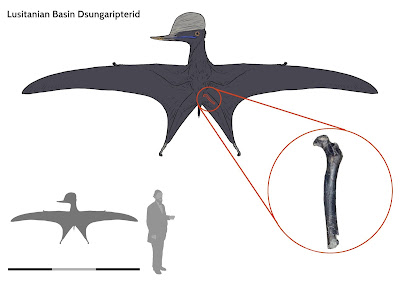
[Paleontology • 2021] A Large Pterosaur Femur (Pterosauria: Dsungaripteroidea) from the Kimmeridgian, Upper Jurassic of Lusitanian Basin, Portugal


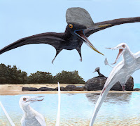
 Pterosaur from the Kimmeridgian, Upper Jurassic of Lusitanian Basin, Portugal

in Bertozzo, Da Silva, Martill, et al., 2021. 
Artwork by Joschua Knüppe twitter.com/JoschuaKnuppe 

The pterosaur fossil record in Portugal is scarce, comprising mainly isolated teeth and rare postcranial material. Here,we describe a well-preserved right proximal femur of a pterodactyloid pterosaur from the Kimmeridgian, Upper Jurassic Praia da Amoreira–Porto Novo Formation of Peniche, Portugal. It is noteworthy for its relatively large size, compared toother Jurassic pterosaurs. It shows affinities with dsungaripteroids based on a combination of features including the bowing of the shaft, the mushroom-like cap of the femoral head, and the distinctly elevated greater trochanter. The femur hasa relatively thinner bone wall compared to dsungaripterids, and is more similar to basal dsungaripteroids. A histological analysis of the bone cortex shows it had reached skeletal maturity. The preserved last growth period indicates fast, uninterrupted growth continued until the final asymptotic size was reached, a growth pattern which could best be compared to pterodactyloid femora from the Early Cretaceous. The specimen is the second confirmed report of a dsungaripteroid from the Jurassic, and it is the first record of this group from the Iberian Peninsula.

Key words: Pterosauria, Dsungaripteroidea, histology, Late Jurassic, Kimmeridgian, Portugal.

A partial proximal right femur of a large indeterminate dsungaripteroid pterosaur (SHN.013) from the Kimmeridgian, Upper Jurassic, Praia da Armoreira-Porto Novo Formation, Peniche, Portugal.
 In ventral (posterior) (A1), dorsal (anterior) (A2), medial (A3), lateral (A4), distal (A3), and proximal (A6) views; A7, drawing of the distal end showing the thickness of the cortex compared to the lumen. Note that the sketch is about twice the scale of the images. The red dashed lines in A2 and A3 indicate the site where the histological section was taken.


Pterosauria Kaup, 1834 
Monofenestrata Lü, Unwin, Jin, Liu, and Ji, 2009 
Pterodactyloidea Plieninger, 1901 

Dsungaripteroidea Young, 1964 (sensu Unwin 2003) 

Dsungaripteroidea indet.

SHN.013


Filippo Bertozzo, Bruno Camilo Da Silva, David Martill, Elsa Marlene Vorderwuelbecke, Tito Aureliano, Remmert Schouten, and Pedro Aquino. 2021. A Large Pterosaur Femur from the Kimmeridgian, Upper Jurassic of Lusitanian Basin, Portugal. Acta Palaeontologica Polonica.  in press. DOI: 10.4202/app.00858.2020

CI2Paleo, Sociedade de Historia Natural, Travessa Florêncio Augusto Chagas nº8B, R/C, 2560-230 Torres Vedras, Portugal.