Sunday, February 25, 2018

[Botany • 2018] Acantholimon ibrahimii • A New Species of A. section Staticopsis (Plumbaginaceae) from the Mediterranean Part of Turkey


Acantholimon ibrahimii  Akaydın

in Akaydın, 2018.

Abstract

A new species, Acantholimon ibrahimii Akaydın, is described, illustrated and discussed in comparison with its close relative A. davisii. The new species is distinguished from the latter species mainly by the generative organs (namely the inflorescence types and petals colour), habitat type and ecological behaviour. Data are also reported on the conservation status of A. ibrahimii, which is suggested to be labelled as EN according to the IUCN categories. Furthermore, a revised key to the Turkish Acantholimon species of A. sect. Staticopsis with spike laxly distichous and scape much longer than leaves is presented.

Keywords: Acantholimon, A. sect. Staticopsis, conservation, endemism, Staticoideae, taxonomy, Eudicots


Galip Akaydın. 2018. Acantholimon ibrahimii (Plumbaginaceae), A New Species of A. section Staticopsis from the Mediterranean Part of Turkey. Phytotaxa. 340(1); 48–54. DOI: 10.11646/phytotaxa.340.1.2

Saturday, February 24, 2018

[Ichthyology • 2018] The Identity of Aplocheilus andamanicus (Köhler, 1906) (Teleostei: Cyprinodontiformes), An Endemic Killifish from the Andaman Islands, with Notes on Odontopsis armata


Aplocheilus andamanicus  (Köhler, 1906) 

in Katwate, Kumkar, Britz, Raghavan & Dahanukar, 2018. 

Abstract

In his work on the fishes of the Andaman Islands, Francis Day (1870) collected large-sized specimens of Aplocheilus from the south Andamans. Despite differences in the size and dorsal-fin ray counts, Day refrained from recognising the Andaman Aplocheilus as a distinct species and considered it as Aplocheilus panchax, a species distributed in the Ganges delta and across the eastern coast of mainland India. However, Day mentioned the differences in fin-ray counts between these two populations. Subsequently Köhler (1906) described the Andaman population as Haplochilus andamanicus (now in Aplocheilus), referring to the diagnostic characters initially discovered by Day. This species failed to receive recognition from taxonomists, because of the uncertainty regarding the validity of the species and its questionable synonymy with A. panchax. In this study, based on morphological and molecular evidence, we demonstrate that A. andamanicus is indeed a distinct and valid species, which can easily be diagnosed from the widespread A. panchax. While resolving the identity of A. andamanicus, we also demonstrate that the congeners from southeast Asia form a genetically distinct group for which the name Odontopsis armata is available.

Keywords: Pisces, Aplocheilus panchax, freshwater fish, taxonomy, South Asia


Unmesh Katwate, Pradeep Kumkar, Ralf Britz, Rajeev Raghavan and Neelesh Dahanukar. 2018. The Identity of Aplocheilus andamanicus (Köhler, 1906) (Teleostei: Cyprinodontiformes), An Endemic Killifish from the Andaman Islands, with Notes on Odontopsis armata van HasseltZootaxa. 4382(1); 159–174.  DOI:  10.11646/zootaxa.4382.1.6


[Botany • 2018] Hyobanche hanekomii • A New Species (Orobanchaceae) from the Western Cape of South Africa


Hyobanche hanekomii  A. Wolfe
  H. atropurpurea Bolus
  H. sanguinea L. 

in Wolfe, 2018. 
DOI: 
10.11646/phytotaxa.340.1.5

Abstract 

The new species Hyobanche hanekomii is described and illustrated. It is somewhat intermediate in appearance between H. sanguinea and H. atropurpurea, but can be distinguished from both in several morphological characters that are presented. The new species occurs in the Cape Fold Belt Mountains of the northwest part of the Western Cape.

 Key words: Cape Floristic Region, holoparasite, parasitic plant


FIGURE 3. Comparison of Hyobanche hanekomii (A), H. atropurpurea (B), and H. sanguinea (C).
Upper panel represents side and front views of corollas to scale (bar = 10 mm).
Lower panel shows inflorescences (not to scale). Illustrations and photographs by A. Wolfe; photos are from living plants in prime blooming condition. Vouchers: (A) upper panel: Hanekom 2887 (1997, NBG); lower panel: Wolfe 1009 (2001, OS); (B) Wolfe 1227 (2006, OS); (C) Wolfe 1387 (2013, OS). 

Hyobanche hanekomii A. Wolfe spec. nov.
. Corolla deep magenta to magenta-red, inflated above the tube and semi-galeate, and 1.5–2.0 times the length of the calyx. 

TYPE:—South Africa. Western Cape: ..., 400 m, 26 September 1997, W.J. Hanekom 2887 (Holotype: NBG 759260!) 

Distribution. Rocky soils in Cape Fold Belt Mountains of northwestern region of the Western Cape, from Citrusdal area to Giftberg (Fig. 2). 

Etymology. The specific epithet is in honour of Mr. Willem Johannes Hanekom (b. 1931). Mr. Hanekom is a keen observer of the flora of the Western Cape, and introduced the author to this new species in 2001. He had made a collection in 1997 (W. J. Hanekom 2887), which included the following note: “Hyobanche sanguinea L. but with influence of H. atropurpurea H. Bol.” 


Andrea D. Wolfe. 2018. Hyobanche hanekomii (Orobanchaceae), A New Species from the Western Cape of South Africa. Phytotaxa. 340(1); 93–97.  DOI: 10.11646/phytotaxa.340.1.5


Friday, February 23, 2018

[Botany • 2018] Rediscovery of Thismia neptunis (Thismiaceae) After 151 Years in the Gunung Matang massif, Borneo


Thismia neptunis Beccari

in Sochor, Egertová, Hroneš & Dančák, 2018. 

Abstract 
Thismia neptunis, as many of its congeners, is a poorly understood species that has only been known from the type collection and its limited original description. In January 2017 it was rediscovered in the type area in the Gunung Matang massif, western Sarawak, Borneo, Malaysia. The paper provides the amended description and drawings of the species, very first available photographs and short notes on taxonomy and historical context of Beccari’s work on Thismia

Key words: Brunonithismia, Burmanniaceae, fairy lanterns, Kubah, Monte Mattán, Sarawak


FIGURE 3. Thismia neptunis: flowering plants (A, B), bud (C), detail of flower (D), section of floral tube and outer view of connective tube (E), detail of inner perianth lobe (F), stigma (G), lateral appendage (H).

Taxonomic treatment 
Thismia neptunis Beccari (1878: 251)

 Type:—MALAYSIA. Ragiato di Sarawak, Mattang. April 1866. O. Beccari p.b. 1508 (holotype FI-B 013453!)

Habitat and ecology:—The only known locality is in primary lowland mixed dipterocarp forest on a river alluvium. Thismia species are generally accompanied by other mycoheterotrophic plants; in this case it was Sciaphila cf. alba Tsukaya & Suetsugu (2015: 284). Albeit pollination ecology was not studied, ca. seven flies of family Sciaridae (Diptera) and one individual of family Braconidae (Hymenoptera) were observed being stuck on inner perianth lobes of the two flowers (Fig. 3A, D, E, F). Although the braconid was probably only a coincidental victim, the flies may represent potential pollinators, as several dipteran taxa have been reported as visitors and probable pollinators of fairy lanterns (Li & Bi 2013, Mar & Saunders 2015). Nevertheless, why had they been attracted to and finally trapped on the perianth lobes surface can only be speculated. Tepals are apparently hydrophilic (possibly as a mean of maintaining turgor in the long thin appendages) as indicated by a number of rain drops persisting on them long after the rain. But they do not appear to be sticky and no other particles tended to be trapped on them either in the field or during our manipulation. Therefore, the insects seem to have been attracted by smell (or other signals) of the flowers and accidentally drowned on the wet surface of perianth lobes.

Distribution:—The species is known from a restricted area in western Sarawak, Borneo, Malaysia. Beccari (1878) described the locality simply as “Monte Mattán” or “Mattang”, which is an area now generally known as Matang massif which Kubah National Park is part of it. The present locality is placed at the park’s western border and may be identical or close to that of Beccari.

Taxonomic affinities:—Having free perianth lobes of unequal length and shape, T. neptunis belongs to section Thismia (Euthismia Schlechter, 1921: 34), subsection Brunonithismia Jonker (1938: 242). This group comprises nine species (Kumar et al. 2017, Suetsugu et al. 2018) of very diverse morphology as for symmetry of perianth, modification of perianth lobes and structure of connectives. Half of the species are, nevertheless, only poorly documented. Thismia neptunis is unique among other fairy lanterns in the very complex three-segmental structure of inner perianth lobes that are terminated by long filiform appendage pointing vertically upwards. This striking morphology led Schlechter to creation of monotypic section Sarawakia Schlechter (1921: 35) within his system of Thismia (Schlechter 1921). However, his approach has not been generally accepted (Jonker 1938, Kumar et al. 2017). 

Beccari was also well aware of morphological uniqueness of T. neptunis. In the protologue (Beccari 1878), he stated that T. neptunis seems to have connectives similar to T. brunonis Griffith (1844: 221). However, T. brunonis have apical part of the connective covered by numerous short teeth (Griffith 1845) while T. neptunis have only three rather long appendages. Nevertheless, Beccari himself was not absolutely sure about the character of connectives as he studied only two pressed and dried plants. In having whitish perianth tube with 12 orange streaks T. neptunis superficially resembles T. javanica Smith (1910: 32) and T. arachnites Ridley (1905: 197). Both of them, nevertheless, differ in having short rounded outer perianth lobes and simpler spreading inner perianth lobes, and the latter species also in having “numerous short teeth” at the apical end of connectives. Connectives of T. javanica, although similar at a first glance, differ from those of T. neptunis in colour (white vs. orange, respectively) and three short teeth at the apex, each bearing 1–2 long hairs of similar length (vs. three unequal filiform appendages in T. neptunis). Thismia neptunis is so far the only known member of subsection Brunonithismia occurring in Borneo.


Michal Sochor, Zuzana Egertová, Michal Hroneš and Martin Dančák. 2018. Rediscovery of Thismia neptunis (Thismiaceae) After 151 Years. Phytotaxa. 340(1); 71–78.  DOI: 10.11646/phytotaxa.340.1.5


[Herpetology • 2018] An Integrative Taxonomic Review of the South Asian Microhylid Genus Uperodon


Uperodon rohani
Garg, Senevirathne, Wijayathilaka, Phuge, Deuti, Manamendra-Arachchi, Meegaskumbura & Biju, 2018


Abstract

Based on a recent molecular phylogenetic study, the South Asian microhylid genus Uperodon (subfamily Microhylinae) currently comprises of 12 valid species that are largely restricted to India and Sri Lanka. Considering the revised generic-level status of its various members, here we review the taxonomy of all known species in this genus and clarify their nomenclatural status and geographical distribution, by integrating evidence from genetics, adult and tadpole morphology, breeding ecology, and bioacoustics. Our molecular analyses of a mitochondrial 16S rRNA gene fragment combined with external and internal morphological studies also revealed a distinct new species in the genus. This species, formally described as Uperodon rohani sp. nov., is endemic to Sri Lanka and widely distributed at lower elevations in the island. For nomenclatural stability of various previously known members, the following actions are also undertaken: (1) redescription of the poorly-defined species Ramanella anamalaiensis Rao (= Uperodon anamalaiensis) and Hylaedactylus montanus Jerdon (= Uperodon montanus); (2) neotype designation for Ramanella anamalaiensis Rao (= Uperodon anamalaiensis), Ramanella minor Rao, Ramanella mormorata Rao (= Uperodon mormorata), and Ramanella triangularis rufeventris Rao; (3) lectotype designation for Callula variegata Stoliczka (= Uperodon variegatus); and (4) synonymization of Ramanella minor Rao with Uperodon anamalaiensis.

Keywords: Amphibia, Amphibians, bioacoustics, endemism, mitochondrial DNA, natural history, neotype, lectotype, new species, tadpoles, Western Ghats-Sri Lanka biodiversity hotspot


Uperodon palmatus (Parker, 1934)


Sonali Garg, Gayani Senevirathne, Nayana Wijayathilaka, Samadhan Phuge, Kaushik Deuti, Kelum Manamendra-Arachchi, Madhava Meegaskumbura and SD Biju. 2018. An Integrative Taxonomic Review of the South Asian Microhylid Genus UperodonZootaxa.  4384(1); 1–88.  DOI:  10.11646/zootaxa.4384.1.1


Thursday, February 22, 2018

[Mammalogy • 2018] Molossus fentoni • A New Species of Mastiff Bat (Chiroptera, Molossidae, Molossus) from Guyana and Ecuador


Molossus fentoni
Loureiro, Lim & Engstrom, 2018


Abstract
We describe a new species of mastiff bat in the genus Molossus (Molossidae), which was previously confused with the common and widely distributed M. molossus, from Guyana and Ecuador based on morphological and molecular differences. It is diagnosed by the following set of morphological characteristics: bicolored dorsal pelage, rounded anterior arch of the atlas, triangular occipital bone, and smaller body and skull size. In a molecular phylogenetic analysis of mitochondrial and nuclear DNA, maximum likelihood and parsimony trees recovered eight clades in the genus and a polyphyletic relationship for the M. molossus species complex. The new species was recovered in a well-supported clade that can be genetically distinguished from other species in the genus by its high level of sequence divergence based on the mitochondrial CO1 gene (8.0–10.1%) and on the nuclear gene beta fibrinogen (1.0–3.1%). It is broadly sympatric with M. molossus sensu stricto in northern South America, but morphologically distinct and genetically divergent.

 Keywords: Molossidae, New species, Phylogenetics, South America, Taxonomy


Fig. 5. Holotype of Molossus fentoni sp. nov. (ROM 122583). Adult male with a medium brown dorsal pelage.

Fig. 4. Dorsal, ventral, posterior, and lateral views of the skull of the holotype of Molossus fentoni sp. nov.

Molossus fentoni sp. nov. 

Diagnosis: A set of traits distinguishes Molossus fentoni from other Molossus. In M. fentoni the infra-orbital foramen is laterally directed; the basioccipital pits are rounded and of moderate depth; the occipital is triangular in posterior view; the upper incisors are thin and long with parallel tips and project forward in an oblique plane relative to the anterior face of the canines (Fig. 4); and the anterior arch of the atlas is rounded (Fig. 6).

Distribution: Molossus fentoni is currently known from the administrative regions of Potaro-Siparuni and Upper Takutu-Upper Essequibo in Guyana and in Orellana province in Ecuador. Although, it has not been documented in the intervening 2000 km of lowland Amazonian forest, we anticipate that it will be found to have a broader distribution then initially represented in our collections. One individual of M. fentoni was collected in syntopy with M. coibensis, M. m. molossus, and M. rufus at ... east of Pompeya Sur, Orellana, Ecuador on 18 May, 2006.

Etymology: This species is named in honour of M. Brock Fenton, Professor Emeritus, Western University, London, Ontario, and one of the world’s foremost researchers in bat ecology and behaviour. He was born in Guyana to Canadian parents and conducted fieldwork in the country in 1970.

Taxonomic remarks: Husson (1962) designated the lectotype of M. molossus as the larger of the two bats described by Buffon and Daubenton (1763). Later, Husson (1962) restricted the type locality of M. molossus to Martinique, which previously had only been designated as the Americas in the first citation of this specimen (Buffon and Daubenton, 1759). Specimens of M. molossus from Martinique were morphologically analyzed in our study and have all the characteristics described above for M. molossus, and not for M. fentoni. In addition, the DNA sample of M. molossus from Martinique clustered with several other samples of M. molossus from the mainland in the phylogenetic trees (Fig. 2), such as Guyana, Suriname, and Brazil, corroborating its affiliation with M. molossus and the distinction from M. fentoni.

Fig. 8. Schematic comparison of cranial features in Molossus.
A and B – Posterior view; C and D – frontal view; E and F – Ventral view. Numbers represent characters described in the text. 1 – Lambdoidal crest and occipital complex; 2 – Sagittal crest; 3 – Mastoid process; 4 – Infra-orbital foramen; 5 – Upper incisors; 6 – Rostrum shape; 7 – Basioccipital pits.


 Livia O. Loureiro, Burton K. Lim and Mark D. Engstrom. 2018. A New Species of Mastiff Bat (Chiroptera, Molossidae, Molossus) from Guyana and Ecuador. Mammalian Biology. 90; 10-21.  DOI: 10.1016/j.mambio.2018.01.008 

[Entomology • 2018] Revision of the Genus Callipia Guenée, 1858 (Lepidoptera, Geometridae), with the Description of 15 New Taxa


Callipia rosetta Thierry-Mieg, 1904
Callipia walterfriedlii  Brehm, 2018
Callipia augustae Brehm, 2018

   DOI:  10.5852/ejt.2018.404 

Abstract

The vividly coloured Neotropical genus Callipia Guenée (1858) (Lepidoptera Linnaeus, 1758, Geometridae (Leach, 1815), Larentiinae (Leach, 1815), Stamnodini Forbes, 1948) is revised and separated into four species groups, according to a provisional phylogeny based on Cytochrome Oxidase I (COI) gene data and morphology. 

Fourteen new species are described using COI data and morphology:
a) in the balteata group: C. fiedleri sp. nov., C. jakobi sp. nov., C. lamasi sp. nov.;
b) in the vicinaria group: C. hausmanni sp. nov., C. walterfriedlii sp. nov.;
c) in the parrhasiata group: C. augustae sp. nov., C. jonai sp. nov., C. karsholti sp. nov., C. levequei sp. nov., C. milleri sp. nov., C. sihvoneni sp. nov., C. wojtusiaki sp. nov. and
d) in the constantinaria group: C. hiltae sp. nov., C. rougeriei sp. nov.
 One new subspecies is described: C. wojtusiaki septentrionalis subsp. nov. 

Two species are revived from synonymy: C. intermedia Dognin, 1914 stat. rev. and C. occulta Warren, 1904 stat. rev. 

The taxon hamaria Sperry, 1951 is transferred from being a junior synonym of C. constantinaria Oberthür, 1881 to being a junior synonym of C. occulta stat. rev. The taxon admirabilis Warren, 1904 is confirmed as being a junior synonym of C. paradisea Thierry-Mieg, 1904. The taxon languescens Warren, 1904 is confirmed as being a junior synonym of C. rosetta, Thierry-Mieg, 1904 and the taxon confluens Warren, 1905 is confirmed as being a junior synonym of C. balteata Warren, 1905. 

The status of the remaining species is not changed: C. aurata Warren, 1904, C. brenemanae Sperry, 1951, C. parrhasiata Guenée, 1858, C. flagrans Warren, 1904, C. fulvida Warren, 1907 and C. vicinaria Dognin. 

All here recognised 26 species are illustrated and the available molecular genetic information of 25 species, including Barcode Index Numbers (BINs) for most of the taxa is provided. The almost threefold increase from 10 to 26 valid species shows that species richness of tropical moths is strongly underestimated even in relatively conspicuous taxa. Callipia occurs from medium to high elevations in wet parts of the tropical and subtropical Andes from Colombia to northern Argentina. The early stages and host plants are still unknown.

Keywords: Callipia; taxonomy; Andes; insect; Neotropics


Figs 131–138. Living specimens and habitats. 131. Callipia rosetta Thierry-Mieg, 1904, ♂, Ecuador, Loja province, Podocarpus National Park, Cajanuma, 2897 m, 26 Mar. 2011. The specimen was attracted to light and benumbed. 132. Elfin forests are a habitat of C. rosetta Thierry-Mieg, 1904 and C. walterfriedlii sp. nov., Ecuador, Loja province, Podocarpus National Park, Cajanuma, 3000 m, 30 Jan. 2013. 133. C. walterfriedlii sp. nov., ♀, Ecuador, Loja province, Podocarpus National Park, Cerro Toledo, 2938 m, 27. Feb. 2013. The specimen was attracted to light and benumbed. 134. Habitat (elfin forest) of C. walterfriedlii sp. nov. at Cerro Toledo. 

Figs 131–138. Living specimens and habitats. 135. Callipia augustae sp. nov., ♂, Peru, Cusco province, Wayqecha station, 2900 m, 26 Aug. 2016. The specimen was collected at night, trapped, photographed and released the next morning. 136. Habitat of C. augustae sp. nov. and Callipia sp. near Wayqecha station. 137. C. augustae sp. nov., ♂, Peru, Cusco province, road Wayqecha–Pillcopata, 2284 m, 23 Aug. 2016. The specimen was attracted to UV light and tried to take up fluid (see proboscis). 138. Callipia sp. at Wayqecha station, 4 Sep. 2016. This specimen was attracted to UV light, but escaped into the vegetation when disturbed.


Gunnar Brehm. 2018. Revision of the Genus Callipia Guenée, 1858 (Lepidoptera, Geometridae), with the Description of 15 New Taxa. European Journal of Taxonomy. 404; 1–54.   DOI:  10.5852/ejt.2018.404

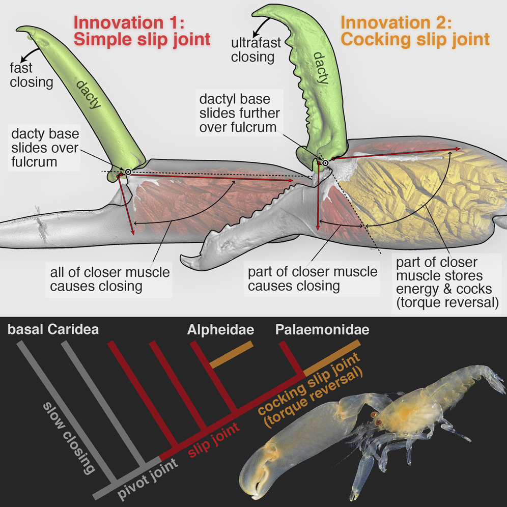
[Crustacea • 2018] Parallel Saltational Evolution of Ultrafast Movements in Snapping Shrimp Claws


Kaji, Anker, Wirkner & Palmer, 2018.

Highlights
• The evolutionary history of remarkable snapping claws in shrimp is reconstructed
• Two novel claw-joint types—slip joints and torque-reversal joints—preceded snapping
• The transition “slip joint → torque-reversal joint → snapping” occurred in two families
• Subtle changes in joint form yielded dramatic changes in claw function (e.g., speed)

Summary
How do stunning functional innovations evolve from unspecialized progenitors? This puzzle is particularly acute for ultrafast movements of appendages in arthropods as diverse as shrimps, stomatopods, insects, and spiders. For example, the spectacular snapping claws of alpheid shrimps close so fast (∼0.5 ms) that jetted water creates a cavitation bubble and an immensely powerful snap upon bubble collapse. Such extreme movements depend on (1) an energy-storage mechanism (e.g., some kind of spring) and (2) a latching mechanism to release stored energy quickly. Clearly, rapid claw closure must have evolved before the ability to snap, but its evolutionary origins are unknown. Unearthing the functional mechanics of transitional stages is therefore essential to understand how such radical novel abilities arise. We reconstructed the evolutionary history of shrimp claw form and function by sampling 114 species from 19 families, including two unrelated families within which snapping evolved independently (Alpheidae and Palaemonidae). Our comparative analyses, using micro-computed tomography (microCT) and confocal imaging, high-speed video, and kinematic experiments with select 3D-printed scale models, revealed a previously unrecognized “slip joint” in non-snapping shrimp claws. This slip joint facilitated the parallel evolution of a novel energy-storage and cocking mechanism—a torque-reversal joint—an apparent precondition for snapping. Remarkably, these key functional transitions between ancestral (simple pinching) and derived (snapping) claws were achieved by minute differences in joint structure. Therefore, subtle changes in form appear to have facilitated wholly novel functional change in a saltational manner.

Keywords: Alpheidae, Palaemonidae, innovation, functional morphology, biomechanics, evolutionary morphology, evo-devo, comparative morphology, saltational evolution, torque-reversal joint


Figure 1. MicroCT Images, Torque Moment Arms, and Schematic Illustrations of Three Shrimp Claw-Joint Types When Closed and Fully Open.
(A) Pivot joint: anterior face∗ of right P1 in a basally branching caridean shrimp. (B) Simple slip joint (no torque reversal or power amplification): anterior face∗ of right P2 in an “intermediate” caridean shrimp. (C) Cocking slip joint (type 1 torque-reversal cocking, most likely power-amplified closing): anterior face∗ of right P1 in a feebly snapping alpheid shrimp. (A’–C’) Overlaid sagittal plane and surface rendering (via micro-computed tomography [microCT]) of claws of all three species showing torque moment-arms (+, –) when closed (upper) and fully opened (lower and background); negative torque (–) indicates that initial contraction of part of the closer muscle causes cocking. (A”–C”) Schematic representation of all three joint types showing loading orientations of opener and closer muscles. (A”) Pivot joint: purely rotational motion of dactyl. (B”) Slip joint: during opening, the dactylar base both rotates and translates (slips) across the propodal ridge (B). (C”) Cocking slip joint: during opening, the dactylar base both rotates and translates—including an abrupt sliding motion into the fully cocked position, where part of the closer muscle (gold) will generate reversed torque (–), and hence energy storage, because it inserts above the fulcrum (white dot).

 White dots show primary rotation axes (A–A”) or fulcrum points (B–B” and C–C”) for dactylar sliding and rotation. Black dots identify a reference point on the dactylar base. White arrows (A–C and A”–C”) show dactylar base trajectories during opening; closing would follow the same trajectories but in reverse. Red arrows (A’–C’) indicate dorsal-most closer-muscle contraction vectors (labeled V1 in Figure 4). Yellow arrows (A’–C’) represent torque moment arms about the fulcrum. Scale bars, 500 μm (A) and 300 μm (B and C). om, opener muscle; cm, closer muscle; (+), positive (counterclockwise) initial torque during claw closing; (–), negative (clockwise) torque during claw cocking generated by the gold-shaded muscle region in (C”). See also Figure SM1 in Methods S1 (joint-type scoring), Figures S1–S4 (microCT images of all claws), Movies S1A and S1B (actual dactyl motion), Movies S2A–S2E (3D model tests), and Table S1 (joint types of all species). ∗See Supplemental Results (Methods S1) for an explanation of claw-face viewing perspectives.


 Tomonari Kaji, Arthur Anker, Christian S. Wirkner and A. Richard Palmer. 2018. Parallel Saltational Evolution of Ultrafast Movements in Snapping Shrimp Claws. Current Biology.  28(1); 106-113.  DOI: 10.1016/j.cub.2017.11.044 

An adaptation 150 million years in the making phy.so/434192683 via @physorg_com

[Crustacea • 2018] Rodriguezia adani • A New Species of Stygobitic Freshwater Crab of the Genus Rodriguezia Bott, 1969 (Decapoda: Trichodactylidae) from Tabasco, Mexico


Rodriguezia adani
Alvarez & Villalobos, 2018


Abstract

A new species of freshwater crab of the family Trichodactylidae, genus Rodriguezia Bott, 1969 is described from Grutas de Agua Blanca in southern Tabasco, Mexico. Rodriguezia is a genus endemic to northern Chiapas and southern Tabasco, distributed over a small area of 70 km. Rodriguezia adani n. sp., the third species of the genus, occurs north of its two congeners, being stygobitic with obvious adaptations to cave life. It can be distinguished from R. villalobosi, an epigean species, by the absence of eyes, lack of pigmentation and elongation of the pereiopods; and from R. mensabak by having less elongated pereiopods relative to carapace breadth, an extremely reduced ocular peduncle, and a smaller adult size.

Keywords: Crustacea, Trichodactylinae, stygobitic, Grutas de Agua Blanca, Tabasco, Chiapas


FIGURE 2. Rodriguezia adani n. sp. male holotype: dorsal view. 

Rodriguezia adani n. sp.

Distribution. The new species is only known from Grutas de Agua Blanca, Macuspana, Tabasco, Mexico.

Etymology. We name the new species after Adán Gómez-González, explorer, biologist and friend, who found these crabs while exploring caves in Tabasco and Chiapas, Mexico.


Fernando Alvarez and José Luis Villalobos. 2018. A New Species of Stygobitic Freshwater Crab of the Genus Rodriguezia Bott, 1969 (Crustacea: Decapoda: Trichodactylidae) from Tabasco, Mexico.  Zootaxa. 4378(1); 137-143. DOI:  10.11646/zootaxa.4378.1.10

Dedican Nueva Especie de Crustáceo al Joven Biólogo Asesinado en Chiapas:"Rodriguezia adani"... - Biosfera 10  biosfera10.org/bios/index.php/noticias/79-nacionales/212-dedican-nueva-especie-de-crustaceo-al-joven-biologo-asesinado-en-chiapas-rodriguezia-adani via @@biosferadiez

[Cnidaria • 2018] A Simple Molecular Technique for Distinguishing Species reveals Frequent Misidentification of Hawaiian Corals in the Genus Pocillopora


colonies of Pocillopora spp. from O‘ahu, Hawai‘i;
(B–D) Pocillopora ligulata(F–I) P. meandrina and (K–M) P. eydouxi

Johnston​, Forsman & Toonen, 2018.
 DOI:  10.7717/peerj.4355 

Abstract
Species within the scleractinian genus Pocillopora Lamarck 1816 exhibit extreme phenotypic plasticity, making identification based on morphology difficult. However, the mitochondrial open reading frame (mtORF) marker provides a useful genetic tool for identification of most species in this genus, with a notable exception of P. eydouxi and P. meandrina. Based on recent genomic work, we present a quick and simple, gel-based restriction fragment length polymorphism (RFLP) method for the identification of all six Pocillopora species occurring in Hawai‘i by amplifying either the mtORF region, a newly discovered histone region, or both, and then using the restriction enzymes targeting diagnostic sequences we unambiguously identify each species. Using this approach, we documented frequent misidentification of Pocillopora species based on colony morphology. We found that P. acuta colonies are frequently mistakenly identified as P. damicornis in Kāne‘ohe Bay, O‘ahu. We also found that P. meandrina likely has a northern range limit in the Northwest Hawaiian Islands, above which P. ligulata was regularly mistaken for P. meandrina.


Figure 3: Images of Pocillopora ligulata colonies, (A)–(E); P. meandrina colonies, (F)–(J); and P. eydouxi colonies, (K)–(O) from O‘ahu, Hawai‘i. 

Figure 1: Pocillopora species composition across the Hawaiian Islands for samples collected from colonies demonstrating P. meandrina morphology. The size of the pie chart is proportional to the number of individuals sampled per island. Pocillopora species are represented by different colors, specifically: P. meandrina, light yellow; P. eydouxi, dark yellow; P. ligulata, light blue; and P. verrucosa, dark blue.

Conclusions: 
Here, we present an assay that allows rapid and unambiguous identification of all six species of Pocillopora present in Hawai‘i, which we hope will work anywhere these species are found. We present two cases where samples identified morphologically were misidentified to highlight the utility of this approach. Taxonomic confusion can impact a wide range of studies and the ability to rapidly and cost-effectively distinguish among species of Pocillopora will benefit future studies of population structure, ecology, biodiversity, evolution and conservation in this challenging genus.


Erika C. Johnston​, Zac H. Forsman and Robert J. Toonen. 2018. A Simple Molecular Technique for Distinguishing Species reveals Frequent Misidentification of Hawaiian Corals in the Genus Pocillopora.  PeerJ. 6:e4355.  DOI:  10.7717/peerj.4355
  

[Arachnida • 2018] There and Back Again: More on the Taxonomy of the Crab Spiders Genus Epicadus (Thomisidae: Stephanopinae)


Epicadus dimidiaster 
Machado, Teixeira & Lise, 2018


 Abstract
The Neotropical crab spider genera Tobias Simon, 1895 and Epicadus Simon, 1895 comprise species with remarkable somatic morphology and confounding taxonomic history. The results of our recent cladistic analysis corroborate and extend preceding taxonomic assumptions in showing that Tobias is a junior synonym of Epicadus. In the present paper the six species recently transferred from Tobias to Epicadus are redescribed. Two new species are described based on both males and females: Epicadus dimidiaster sp. nov. and Epicadus tigrinus sp. nov.; the male of Epicadus granulatus Banks, 1909 is described for the first time. The diagnosis of the genus is revised, an identification key is provided, and information on geographical distribution is updated. Epicadus now comprises eleven species.

Keywords: Araneae, Dionycha, Neotropical region, Stephanopinae, taxonomy


Miguel Machado, Renato Augusto Teixeira and Arno Antonio Lise. 2018. There and Back Again: More on the Taxonomy of the Crab Spiders Genus Epicadus (Thomisidae: Stephanopinae). Zootaxa. 4382(3);  501–530.  DOI:  10.11646/zootaxa.4382.3.4
Thiago Da Silva Moreira and Miguel Machado. 2016. Taxonomic Revision of the Crab Spider Genus Epicadus Simon, 1895 (Arachnida: Araneae: Thomisidae) with Notes on Related Genera of Stephanopinae Simon, 1895. Zootaxa. 4147(3); 281–310.  DOI: 10.11646/zootaxa.4147.3.4


Wednesday, February 21, 2018

[Botany • 2018] Lecanorchis sarawakensis • A New Mycoheterotrophic Species (Orchidaceae, Vanilloideae) from Sarawak, Borneo


Lecanorchis sarawakensis Suetsugu & Naiki

in  Suetsugu, Ling, Naiki, et al., 2018.

Lecanorchis Blume (1856: 188) comprises about 30 species of mycoheterotrophic orchids (Seidenfaden 1978, Hashimoto 1990, Szlachetko & Mytnik 2000, Govaerts et al. 2017) characterized by having numerous, long, thick, horizontal roots produced from a short rhizome, presence of a calyculus (i.e. a cup-like structure located between the base of the perianth and apex of the ovary) and an elongate column with a pair of small wings on each side of the anther (Seidenfaden 1978, Hashimoto 1990). The genus is distributed across a wide area including China, Korea, India, Indonesia, Japan, Laos, Malaysia, New Guinea, Pacific islands, the Philippines, Taiwan, Thailand and Vietnam (Seidenfaden 1978, Hashimoto 1990, Pearce & Cribb 1999, Szlachetko & Mytnik 2000, Averyanov 2011, 2013).

Lecanorchis sarawakensis in the type locality.
A. Habit. B. Flower, side view. C. Flower, front view.

Lecanorchis sarawakensis Suetsugu & Naiki, sp. nov.


Kenji Suetsugu, Ling Chea Yiing, Akiyo , Shuichiro Tagane, Yayoi Takeuchi, Hironori Toyama and Tetsukazu Yahara. 2018. Lecanorchis sarawakensis (Orchidaceae, Vanilloideae), A New Mycoheterotrophic Species from Sarawak, Borneo. Phytotaxa. 388(1); 135–139. DOI:  10.11646/phytotaxa.338.1.13