Showing posts with label Nature Communications. Show all posts

Sunday, October 11, 2026

[Paleontology • 2026] Infernogallus jamesi • Fruit-eating Adaptations in a Late Cretaceous oviraptorosaur (Oviraptorosauria: Caenagnathidae)


 Infernogallus jamesi 
Funston, Dudgeon, Simon, Dufault, Boyd, Fowler, Currie & Evans, 2026
 

Abstract
The diversification of herbivorous dinosaurs and the rise of angiosperms in the Late Cretaceous are each well established, but the link between them is debated. Broad-scale diversity patterns in the two groups appear not to closely align, and there is no direct evidence of dinosaur adaptation to angiosperm-based diets. Here we describe Infernogallus jamesi gen. et. sp. nov., a diminutive caenagnathid oviraptorosaur, and show that these theropods were specialised herbivores that originated alongside the rise of fleshy angiosperm fruits. The nearly complete, articulated skull of Infernogallus jamesi—the first for the clade—reveals drastic changes to the adductor musculature organisation, unique in non-avian theropods but converged upon by extant frugivorous parrots. These modifications enabled extensive oral processing of soft food using anteroposterior movement of the mandible and occlusion of ridges and grooves on opposed elements. Altogether, this attests to a highly specialised herbivorous diet in caenagnathids, which likely consisted in large part of fleshy angiosperm fruits, which increased in size and diversity in concert with the radiation of oviraptorosaurs. Caenagnathids thus provide one of the strongest candidates for angiosperm-based diet adaptations in dinosaurs, but their role in the Late Cretaceous proliferation of fruit—which shaped early Cenozoic flora—remains unclear.


Systematic palaeontology
Theropoda
Oviraptorosauria
Caenagnathidae

Eucaenagnathinae Clade nov.

Definition. The most inclusive monophyletic group including Anzu wyliei and Elmisaurus rarus, but not Anomalipes zhaoi.

Diagnosis. Eucaenagnathines (meaning ‘true caenagnathids’) are distinguished from other caenagnathids by the morphology of the fused dentaries, with an upturned tomial edge marked with lateral and anterior occlusal ridges, and a midline sulcus bordered by longitudinal lingual ridges separated from the tomial edge by a lingual groove.

Infernogallus jamesi (ROM VP 65050). Photograph (a) and line drawing (b) of ROM VP 65050. c Skeletal reconstruction of Infernogallus jamesi based on ROM VP 65050 and other eucaenagnathines (shaded areas are preserved in ROM 65050). d Palaeohistological thin section of the femur of ROM VP 65050, showing lines of arrested growth (arrows) and an external fundamental system indicative of advanced age and skeletal maturity. cv cervical vertebrae, dv dorsal vertebrae, efs external fundamental system, fe femur, ga gastralia, hu humerus, il ilium, ma manus, pb pubis, pp plane-polarised light, sc scapulacoracoid, sk skull, tb tibia, xp cross-polarised light. Scale bars as indicated.


Cranial anatomy of Infernogallus jamesi gen. et sp. nov.
Photograph (a) and line drawing (b) of the skull of ROM VP 65050 in left lateral view. ...

 Infernogallus jamesi gen et. sp. nov. (Figs. 1–4) 

Etymology. The name comes from the Latin “Inferno” (Hell) and “gallus” (chicken), referring to the Hell Creek Formation and the small, bird-like appearance of the species; “jamesi”, a patronym for James William Boyd, the paternal grandfather of author CAB, who sparked his interest in the field of palaeontology. 

Holotype. ROM VP 65050, articulated partial skeleton including the skull, cervical and dorsal vertebrae, right pectoral girdle and forelimb, partial pelvic girdle, and partial right hindlimb. Paratypes. ROM VP 64193, dentaries; ROM VP 64194, articularsurangular-coronoid complex; ROM VP 88690, left and right pterygoids; NDGS 4351, dentaries. 


Locality and horizon. Hell Creek Formation (late Maastrichtian), Garfield County, Montana; Meade County, South Dakota; and Bowman County, North Dakota, USA. 


Diagnosis. Small (femur length 185 mm; est. 12 ± 3 kg body mass) caenagnathid oviraptorosaur diagnosed by the following autapomorphies: broad jugal with large anterior ascending process and which contacts the squamosal posteriorly; infratemporal fenestra extremely reduced, about 25% the diameter of the orbit; quadratojugal with greatly enlarged, circular posterior process; squamosal with dorsoventrally broad body lacking a depression and ridge for adductor musculature; pterygoids concave dorsally, unfused along the midline, and with a lateral buttress; ectopterygoid–pterygoid bar greatly expanded and extending far ventrally; long axis of vomer oriented ...


Gregory F. Funston, Thomas W. Dudgeon, D. Jade Simon, Danielle M. Dufault, Clint Boyd, Denver Fowler, Philip J. Currie and David C. Evans. 2026. Fruit-eating Adaptations in a Late Cretaceous oviraptorosaur (Theropoda). Nature Communications. DOI: doi.org/10.1038/s41467-026-78343-w [10 October 2026] 


Thursday, October 1, 2026

[Paleontology • 2026] Norellraptor barsboldi • Independent Assembly of the Flight Apparatus in A Non-avian Dinosaur Clade


 Norellraptor barsboldi 
Wang, Ji, Cau, Kundrát, Liu, Wang & Ji, 2026


Abstract
The discovery of feathered dinosaurs revolutionized the studies on the origin of birds and the evolution of their flight. Among paravians (birds and their closest relatives), the microraptorine dromaeosaurids are unique in bearing several flight-related adaptations with a controversial evolutionary interpretation, supporting, alternatively, flight as ancestral to Paraves or having evolved multiple times among the latter. Here, we introduce an exquisitely-preserved member of Microraptorinae, Norellraptor barsboldi gen. et sp. nov., and describe in detail forelimb osteohistological features for that clade. Norellraptor nests among late-diverging microraptorines and supports step-wise assembly of aerial adaptations in that clade. About 30% of the microraptorine synapomorphies are convergently acquired by the bird lineage (Avialae). Yet, the sequences of flight-related novelties reconstructed along the two lineages differ consistently; combined with histological evidence which might indicate a peculiar limb growth model in Microraptorinae, these results dismiss a shared developmental pattern driving the evolution of the flying dinosaurs and support multiple and independent selective regimes at the origin of the winged taxa in Paraves.


 Norellraptor barsboldi 130108-MHGU-F4281
a Whole specimen. b Skull in lateral view. c Close up of the rostrum. d Close up of the posterior part of skull. e Pectoral region. f Distal end of tail.
aof, antorbital fossa; arm, ascending ramus of maxilla; at, anterior tympanic recess; bt, basal tuber; bp, basypterygoid process; cf, coracoid fenestra; ct, caudal tympanic recess; cnV, 5th cranial nerve opening; cnVII, 7th cranial nerve opening; cv, cervical vertebra; d1, first dorsal vertebra; de, dentary; dt, dorsal tympanic recess; fo, foramen; fr, frontal; fu, furcula; gl, glenoid; h, humerus; la, lacrimal; lat, laterosphenoid; lcv, last caudal vertebra; lp, lateral pneumatic recess; mc, metacarpus; mxf, maxillary fenestra; na, nasal; oc, otosphenoid crest; or, otic region; pa, parietal; paw, postantral wall; pmx, premaxilla; pp, paroccipital process; ppd, posterior processes of dentary; pt, pterygoid; pxf, promaxillary fenestra; r, radius; rp, retroarticular process; sa, surangular; sc, scapula; so, supraoccipital; sp, sternal plate; sr, sclerotic ring; u, ulna; up, uncinate process; vor, ventral otic recess. 
Arrow indicates point of histological sampling. 
Scale bar = 50 mm (a); 10 mm (b–f).


 Norellraptor barsboldi 130108-MHGU-F4281, appendicular elements.
a Right hand. b Pubis. c Feet.
am, acetabular margin; ap, apron; cs, claw soft tissue; gi, gastral impression; mcI-III, metacarpals I to III; mtI-V, metatarsals I to V; p, phalanx; pf, pubic foot; r, radius; sc, semilunate carpal; t, tibiotarsus; u, ulna; uI-III, unguals 1 to 3. 
Scale bar = 10 mm.

Systematic palaeontology
Dinosauria Owen, 1842 
Theropoda Marsh, 1881 

Dromaeosauridae Matthew and Brown, 1922  
Microraptorinae Senter et al., 2004 
Microraptorini (Senter et al., 2004) 

Norellraptor barsboldi gen. et sp. nov.

Locality and horizon: Lamadong Town, Jianchang County, western Liaoning, China. Jiufotang Formation, Lower Cretaceous. China.

Diagnosis: Microraptorine dromaeosaurid with the unique combination of features (autapomorphies marked by asterisk; Supplementary Fig. 2): premaxilla with length of prenarial part half the depth of the subnarial body; large promaxillary fenestra occupying the whole rostral third of the antorbital fossa*, and with long axis inclined 35° caudodorsally; pila promaxillaris narrower than the pila interfenestralis and both less than half the diameter of the maxillary fenestra*; pubis gently curved caudoventrally with small foot having the posterior projection less than the diameter of the pubic shaft*. Differential diagnosis: Norellraptor differs from Changyuraptor in having basally-constricted dentary crowns and in the semilunate carpal barely
 

Etymology: The genus name refers to the American palaeontologist Mark Allen Norell (1957-2025), with Latin word “raptor” (“robber”), a frequently used suffix in dromaeosaurid taxonomy. The species name refers to the Mongolian palaeontologist Rinchen Barsbold (1935-2025). The new taxon name remarks and honours the exceptional contributions by R. Barsbold and M. A. Norell to the study of the bird-like dinosaurs.
 

Xuri Wang, Yannan Ji, Andrea Cau, Martin Kundrát, Yichuan Liu, Yang Wang and Qiang Ji. 2026. Independent Assembly of the Flight Apparatus in A Non-avian Dinosaur Clade. Nature Communications. 17: 10074. DOI: doi.org/10.1038/s41467-026-77804-6 [29 September 2026]

Wednesday, September 24, 2025

[Paleontology • 2025] Joaquinraptor casali • Latest Cretaceous megaraptorid theropod Dinosaur sheds light on megaraptoran evolution and palaeobiology

 
Joaquinraptor casali 
Ibiricu, Lamanna, Alvarez, Cerda, Caglianone, Cardozo, Luna & Martínez, 2025 

Artwork by  Andrew McAfee

Abstract
Recent fossil discoveries have cast considerable light on the palaeobiology of Megaraptora, a group of large-clawed carnivorous theropod dinosaurs known from Cretaceous deposits in Asia, Australia, and especially South America. Nevertheless, many important aspects of megaraptoran morphology and evolution remain poorly understood, due in large part to the fragmentary nature of most fossils of these theropods and the scarcity of anatomically overlapping skeletal elements among the known taxa. Here we report a previously unknown megaraptoran genus and species represented by a partially articulated partial skeleton recovered from an uppermost Cretaceous stratum of the Lago Colhué Huapi Formation of south-central Chubut Province in central Patagonia, Argentina. Pertaining to the derived megaraptoran subclade Megaraptoridae, the taxon is among the most completely represented and latest-surviving megaraptorans. Its stratigraphic occurrence indicates that these dinosaurs likely persisted to the Cretaceous/Palaeogene boundary; moreover, the preservation of a crocodyliform humerus between the dentaries of the new theropod may provide information on megaraptoran dietary preferences and feeding strategies. Megaraptorids appear to have been the apex predators in central and southern Patagonian palaeoecosystems approaching the end of the Cretaceous, in contrast to more northerly areas of South America where these niches were occupied by other non-avian theropod groups.


 Osteology of Joaquinraptor casali gen. et sp. nov.
a Skeletal reconstruction of Joaquinraptor in left lateral view with preserved elements in blue (some reversed from right side) (modified and updated from Lamanna et al.7 [these authors’ Fig. 1e], which was in turn modified by A. McAfee from an original illustration by T.K. Robinson). Right maxilla in lateral (b) and medial (c) views. Skull roof, braincase, and atlantal intercentrum in dorsal (d) and ventral (e) views. f Probable right postorbital in lateral view. g Right quadrate in anterior view. Right and left dentaries in lateral and medial views (right dentary in lateral view and left dentary in medial view in h; opposite in i). j Two articulated middle or posterior caudal vertebrae in right lateral view. k Dorsal rib in anterior view. l Anterior haemal arch in anterior view. m Left scapulocoracoid in lateral view. Left humerus in anterolateral (n), lateral (o), and medial (p) views. Left radius in anterior (q) and lateral (r) views. Left ulna in lateral (s) and posterior (t) views. u Right manual ungual II (=manual phalanx II-3) in lateral view. Left femur in anterior (v) and posterior (w) views. x Distal right tibia in anterior view. y Right pedal ungual III (=pedal phalanx III-4) in medial view. Dashed lines indicate missing areas of scapulocoracoid and femur.

Systematic palaeontology
Saurischia Seeley, 1887 
Theropoda Marsh, 1881 
Tetanurae Gauthier, 1986 

Megaraptora Benson, Carrano, and Brusatte, 2010 
Megaraptoridae Novas, Agnolín, Ezcurra, Porfiri, and Canale, 2013 

Joaquinraptor casali gen. et sp. nov.

Etymology: Joaquín, in tribute to the son of the first author (L.M.I.) and the informal name given to the locality when the skeleton of the taxon was discovered (Valle Joaquín); Latin, raptor, thief. Specific epithet casali in recognition of Dr. Gabriel Andrés Casal for his many contributions to knowledge of the Cretaceous palaeontology and geology of central Patagonia (including the formal recognition and naming of the formation from which this megaraptorid was recovered).


Joaquinraptor casali with an ancient crocodile relative's front leg in its mouth.
Artwork by  Andrew McAfee

 
Lucio M. Ibiricu, Matthew C. Lamanna, Bruno N. Alvarez, Ignacio A. Cerda, Julieta L. Caglianone, Noelia V. Cardozo, Marcelo Luna and Rubén D. Martínez. 2025. Latest Cretaceous megaraptorid theropod Dinosaur sheds light on megaraptoran evolution and palaeobiology. Nature Communications. 16: 8298. DOI: doi.org/10.1038/s41467-025-63793-5 [23 September 2025]


Saturday, December 21, 2024

[Paleontology • 2024] Early–middle Permian Mediterranean gorgonopsian suggests an equatorial origin of therapsids

a gorgonopsian from the island of Mallorca, western Mediterranean

in Matamales-Andreu, Kammerer, Angielczyk, Simões, Mujal, Galobart et Fortuny, 2024.  
Reconstruction by Henry Sutherland Sharpe

Abstract
Therapsids were a dominant component of middle–late Permian terrestrial ecosystems worldwide, eventually giving rise to mammals during the early Mesozoic. However, little is currently known about the time and place of origin of Therapsida. Here we describe a definitive therapsid from the lower–?middle Permian palaeotropics, a partial skeleton of a gorgonopsian from the island of Mallorca, western Mediterranean. This specimen represents, to our knowledge, the oldest gorgonopsian record worldwide, and possibly the oldest known therapsid. Using emerging relaxed clock models, we provide a quantitative timeline for the origin and early diversification of therapsids, indicating a long ghost lineage leading to the evolutionary radiation of all major therapsid clades within less than 10 Myr, in the aftermath of Olson’s Extinction. Our findings place this unambiguous early therapsid in an ancient summer wet biome of equatorial Pangaea, thus suggesting that the group originated in tropical rather than temperate regions.

Systematic palaeontology
Synapsida Osborn, 190313
Therapsida Broom, 190514

Gorgonopsia Seeley, 189415
Gorgonopsia indet.

Most relevant elements of DA21/17-01-01 and silhouette showing their positions


 Life reconstruction of the gorgonopsian from Mallorca in a floodplain setting.
Reconstruction by Henry Sutherland Sharpe


 Rafel Matamales-Andreu, Christian F. Kammerer, Kenneth D. Angielczyk, Tiago R. Simões, Eudald Mujal, Àngel Galobart and Josep Fortuny. 2024. Early–middle Permian Mediterranean gorgonopsian suggests an equatorial origin of therapsids. Nature Communications. 15: 10346. DOI: doi.org/10.1038/s41467-024-54425-5  

Friday, November 1, 2024

[PaleoIchthyology • 2024] Ngamugawi wirngarri • A Late Devonian coelacanth reconfigures Actinistian Phylogeny, Disparity, and Evolutionary Ddynamics

 
Ngamugawi wirngarri 
Clement, Cloutier, Lee, King, Vanhaesebroucke, Bradshaw, Dutel, Trinajstic & Long, 2024 


Abstract
The living coelacanth Latimeria (Sarcopterygii: Actinistia) is an iconic, so-called ‘living fossil’ within one of the most apparently morphologically conservative vertebrate groups. We describe a new, 3-D preserved coelacanth from the Late Devonian Gogo Formation in Western Australia. We assemble a comprehensive analysis of the group to assess the phylogeny, evolutionary rates, and morphological disparity of all coelacanths. We reveal a major shift in morphological disparity between Devonian and post-Devonian coelacanths. The newly described fossil fish fills a critical transitional stage in coelacanth disparity and evolution. Since the mid-Cretaceous, discrete character changes (representing major morphological innovations) have essentially ceased, while meristic and continuous characters have continued to evolve within coelacanths. Considering a range of putative environmental drivers, tectonic activity best explains variation in the rates of coelacanth evolution.


Ngamugawi wirngarri 
A, B ‘Part a’ of WAM 09.6.148 (holotype) shown in left dorsolateral view and skull close up in left lateral view. C ‘Part b’ of WAM 09.6.148 (holotype) showing all exposed elements; D partial braincase of NMV P231504 (paratype) shown in right lateral view; E cleithrum of NMV P231504 (paratype) in mesial and lateral view; F, G skull reconstruction in dorsal and left lateral view.
Abbreviations: Ang angular, Cl cleithrum, Clv clavicle, Dt dentary, Exc extracleithrum, icj intracranial joint, ioc infraorbital canal, L.Gu lateral gular, Lj lachrymojugal, mc mandibular canal, L.Ex lateral extrascapular, Op operculum, Par Parietal, Po postorbital, Pop preoperculum, Pp postparietal, Pmx premaxilla, Psym parasymphysial, Q quadrate, Ro.p1 anterior pore of the rostral organ, Ro.p2 antero-lateral pore of the rostral organ, Ro.p3 postero-lateral pore of the rostral organ, So supraorbitals, soc supraorbital canal, Sop Suboperculum, Spl splenial, Sq squamosal.

Systematic palaeontology
Osteichthyes Huxley 1880
Sarcopterygii Romer 1955
Actinistia Cope 1871

Ngamugawi wirngarri gen. et sp. nov.

Diagnosis: Ngamugawi wirngarri gen. et sp. nov. is distinguished from all other coelacanths by the following apomorphies: jugal canal with prominent branches; large sensory pore openings between supraorbitals and parietals; teeth on parasymphysial tooth plate, but not on the dentary; prearticular and/or coronoid teeth rounded; cleithra and extracleithra with broad triangular anteroventral overlap for clavicle bearing a large ventral foramen; and scales with long ornamental ridges extending beyond the posterior margin of the base (Figs. 1, 2, Supplementary Fig. 1).

Locality and horizon: Canning Basin, in northern Western Australia, circa 100 km southeast of Fitzroy Crossing; Gogo Formation, early Frasnian, Late Devonian (~384–382 Ma). The holotype was found between Stromatoporoid Camp and Longs Well, the paratype was found in Paddys Valley. 

Etymology: Generic name meaning “ancient fish” in Gooniyandi/Guniyandi, language of the First Nations people from Country around Fitzroy Crossing in the Kimberley region of Western Australia. Specific name is given in honour of respected Gooniyandi elder and ancestor Wirngarri, who lived in the Emanuel Range. Generic and specific names were both provided to Prof. John Long in September 2023, who has a longstanding and ongoing relationship with the community, with permissions to use the language granted by elder Rosemary Nuggett, on behalf of the Gooniyandi people of the Mimbi community.


Phylogenetic relationships and divergence dates within coelacanths, based on tip-dated Bayesian inference.

A live recreation of the Ngamugawi wirngarri coelacanth in its natural habitat. P3D graphic credit: Katrina Kenny. 
 Illustration by Katrina Kenny (courtesy Flinders University)


 Alice M. Clement, Richard Cloutier, Michael S. Y. Lee, Benedict King, Olivia Vanhaesebroucke, Corey J. A. Bradshaw, Hugo Dutel, Kate Trinajstic and John A. Long. 2024. A Late Devonian coelacanth reconfigures Actinistian Phylogeny, Disparity, and Evolutionary Ddynamics. Nature Communications. 15: 7529. DOI: doi.org/10.1038/s41467-024-51238-4


Saturday, January 27, 2024

[PaleoEntomology • 2024] Eunotalia emeryi, Cretotettigarcta shcherbakovi & Pranwanna xiai • Mesozoic Evolution of Cicadas and their Origins of Vocalization and Root Feeding


Life reconstruction of cicadas in a Mesozoic Forest. 
Eunotalia emeryi gen. et sp. nov.
Cretotettigarcta shcherbakovi 
sp. nov.
Pranwanna xiai gen. et sp. nov.

 Jiang, Szwedo, Labandeira, Chen, Moulds, Mähler, Muscente, Zhuo, Nyunt, Zhang, Wei, Rust & Wang, 2024
 Reconstructed by Mr. Dinghua Yang

Abstract
Extant cicada (Hemiptera: Cicadoidea) includes widely distributed Cicadidae and relictual Tettigarctidae, with fossils ascribed to these two groups based on several distinct, minimally varying morphological differences that define their extant counterparts. However, directly assigning Mesozoic fossils to modern taxa may overlook the role of unique and transitional features provided by fossils in tracking their early evolutionary paths. Here, based on adult and nymphal fossils from mid-Cretaceous Kachin amber of Myanmar, we explore the phylogenetic relationships and morphological disparities of fossil and extant cicadoids. Our results suggest that Cicadidae and Tettigarctidae might have diverged at or by the Middle Jurassic, with morphological evolution possibly shaped by host plant changes. The discovery of tymbal structures and anatomical analysis of adult fossils indicate that mid-Cretaceous cicadas were silent as modern Tettigarctidae or could have produced faint tymbal-related sounds. The discovery of final-instar nymphal and exuviae cicadoid fossils with fossorial forelegs and piercing-sucking mouthparts indicates that they had most likely adopted a subterranean lifestyle by the mid-Cretaceous, occupying the ecological niche of underground feeding on root. Our study traces the morphological, behavioral, and ecological evolution of Cicadoidea from the Mesozoic, emphasizing their adaptive traits and interactions with their living environments.

Adults, final instar nymph, and exuviae of Cicadoidea fossils in Kachin amber of northern Myanmar.
 a Eunotalia emeryi gen. et sp. nov. (MGM2016–014). This image was published in the study by ref. 41 (Fig. 3a). b Cretotettigarcta problematica comb. nov. (new material: NIGP201895). c Cretotettigarcta shcherbakovi sp. nov. (NIGP201896). d Vetuprosbole parallelica (new material: NIGP201897).
e–i Pranwanna xiai gen. et sp. nov. (LYU–BC2001, male; LYU–BC2002, female). e Dorsal view of male. f Dorsal view of female. g Ventral view of male. h Ventral view of female. i Left view of final-instar nymph, Cicadoidea species 1 (NIGP2018985).
j–m Final- nymphal exuviae. j Nymphal sp. 2 (MGM2016–017), left view. k Nymphal sp. 3 (LYU–BC2004), right view. l Nymphal sp. 4 (NIGP201900), ventral view. m Nymphal sp. 5 (NIGP201901), left view.

Stem cicadoids
Eunotalia gen. nov.
  Eunotalia emeryi sp. nov.  

Etymology: The generic name is a compound form, from Classical Greek prefix: eu-, meaning ‘true’ or ‘good’, and notos, meaning ‘back’ or ‘dorsum’.

Stem cicadids
Cretotettigarcta

Cretotettigarcta problematica comb. nov.
Cretotettigarcta shcherbakovi sp. nov.

Pranwanna gen. nov 
 Pranwanna xiai sp. nov.

Etymology: The generic name, pranwanna, is from the Jingpho language spoken in Kachin State of Myanmar, meaning ‘primitive’.


Hui Jiang, Jacek Szwedo, Conrad C. Labandeira, Jun Chen, Maxwell S. Moulds, Bastian Mähler, A. Drew Muscente, De Zhuo, Thet Tin Nyunt, Haichun Zhang, Cong Wei, Jes Rust and Bo Wang. 2024. Mesozoic Evolution of Cicadas and their Origins of Vocalization and Root Feeding. Nature Communications. 15, 376.  DOI: 10.1038/s41467-023-44446-x

Thursday, November 2, 2023

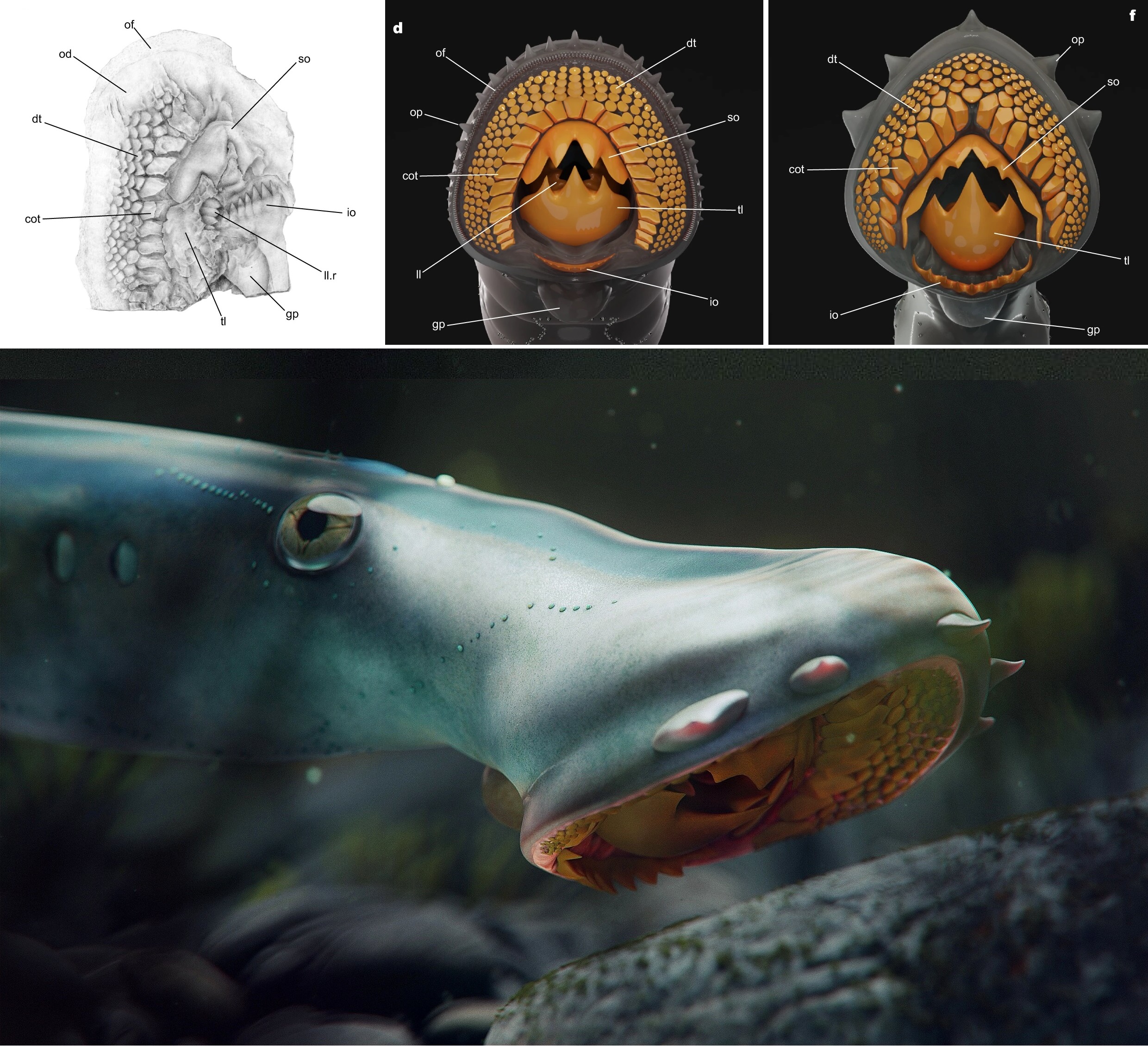
[PaleoIchthyology • 2023] Yanliaomyzon ingensdentes & Y. occisor • The Rise of Predation in Jurassic Lampreys


Yanliaomyzon occisor & Y. ingensdentes 
 Wu, Janvier & Zhang, 2023


Abstract
Lampreys, one of two living lineages of jawless vertebrates, are always intriguing for their feeding behavior via the toothed suctorial disc and life cycle comprising the ammocoete, metamorphic, and adult stages. However, they left a meager fossil record, and their evolutionary history remains elusive. Here we report two superbly preserved large lampreys from the Middle-Late Jurassic Yanliao Biota of North China and update the interpretations of the evolution of the feeding apparatus, the life cycle, and the historic biogeography of the group. These fossil lampreys’ extensively toothed feeding apparatus differs radically from that of their Paleozoic kin but surprisingly resembles the Southern Hemisphere pouched lamprey, which foreshadows an ancestral flesh-eating habit for modern lampreys. Based on the revised petromyzontiform timetree, we argued that modern lampreys’ three-staged life cycle might not be established until the Jurassic when they evolved enhanced feeding structures, increased body size and encountered more penetrable host groups. Our study also places modern lampreys’ origin in the Southern Hemisphere of the Late Cretaceous, followed by an early Cenozoic anti-tropical disjunction in distribution, hence challenging the conventional wisdom of their biogeographical pattern arising from a post-Cretaceous origin in the Northern Hemisphere or the Pangean fragmentation in the Early Mesozoic.

Jurassic lampreys from the Yanliao Biota, China, Yanliaomyzon occisor gen. et sp. nov. and Yanliaomyzon ingensdentes gen. et sp. nov.
a–e Yanliaomyzon occisor gen. et sp. nov., a Photograph of holotype (IVPP V 15830); b Line drawing of the oral disc and dentition of (a), based on Supplementary Fig. 2k and l; c, d Paratype (IVPP V 18956B), photograph (c) and line drawing (d); e Restoration.
 f–h Yanliaomyzon ingensdentes gen. et sp. nov., f Photograph of holotype (IVPP V 16715B), white arrow pointing to the skeletal relics in gut content; g Oral disc and dentition; h Restoration.
Abbreviations: adf, ‘anterior dorsal fin’ (dorsal fin); af, anal fin fold; ba, branchial apparatus; ca, cloaca (anus); cot, circumoral teeth; da, dorsal aorta; dcf, dorsal lobe of caudal fin; dt, oral disc teeth; cf, caudal fin; e, eyes; dt, disc teeth; go, external gill openings; gp, gular pouch; ic, intestine contents; io, infraoral lamina; ll, longitudinal lingual lamina; ll.l, left longitudinal lingual lamina; ll.r, right longitudinal lingual lamina; lv, liver; ns, olfactory organ (nasal sac); oc, otic capsule; od, oral disc; of, oral fimbriae; op, oral papilla(e); paf, precloacal skin fold; pdf, ‘posterior dorsal fin’ (anterior part of caudal fin); pt, piston cartilage; so, supraoral lamina; tl, transverse lingual lamina; vcf, ventral lobe of caudal fin; V1?, ophthalmic ramus of trigeminal nerve?

a–d Oral disc and dentition of Yanliaomyzon ingensdentes gen. et sp. nov., a Photograph (IVPP V 16716B) and b Line drawing; c Photograph (IVPP V 16716A), whitened with ammonium chloride, the white arrow pointing to the imprints of the wrinkles of the gular pouch; d Restoration.
e, f Oral disc and dentition of Yanliaomyzon occisor gen. et sp. nov., e Photograph (IVPP V18956A), whitened with ammonium chloride; f Restoration;
 g Oral disc and dentition of Geotria australis, redrawn from ref. 10.
Abbreviations: cot, circumoral teeth; dt, oral disc teeth; gp, gular pouch; ic, intestine contents; io, infraoral lamina; ll, longitudinal lingual lamina; ll.r, right longitudinal lingual lamina; od, oral disc; of, oral fimbriae; op, oral papilla(e); so, supraoral lamina; tl, transverse lingual lamina.


Systematic paleontology
Order: Petromyzontiformes Berg, 194018

Genus Yanliaomyzon gen. nov.
 
Diagnosis: Stem lampreys with oral discs well-toothed in anterior and lateral fields; anterior and lateral oral disc teeth closely arranged, dorsally truncated, spatulate in shape with the slightly concaved undersurface of the free edge protruding a shallow blade; posterior disc teeth lacking, anterior and lateral circumoral teeth elongate and trihedral in shape; supraoral lamina large and consisting of two stout central cusps flanked by wing-like lateral extensions; transverse lingual lamina very large with the apices of three cusps interlocking with the supraoral lamina in vivo.
 
Etymology: ‘Yanliao’ derives from Yanliao Biota, a Jurassic terrestrial Lagerstätte from North China, where these fossils were discovered; ‘myzon’ (Greek), sucker.

Yanliaomyzon occisor

Diagnosis: The supraoral lamina spanning completely the lateral rims of the oral aperture, with the central cusps flanked immediately by two smaller projections; 16 circumoral teeth; the tail region occupying slightly less than 28% of the total body length.

Etymology: Latin ‘occisor’, meaning ‘killer’, refers to the powerful hunting skill of the species.

Horizon and locality: Tiaojishan Formation, Oxfordian, earliest Late Jurassic, ca. 158.58–160 million years ago (Ma); Daxishan, Linglongta Town, Jianchang County, Liaoning Province (Holotype), and Nanshimen Village, Gangou Town, Qinglong County, Hebei Province (Paratype), China.


Yanliaomyzon ingensdentes gen. et sp. nov.

Diagnosis: The supraoral lamina occupying roughly one-third of the rim of the oral aperture; the transverse lingual lamina almost equaling to the supraoral lamina in width; ca. 23 circumoral teeth; the tail region occupying slightly more than 40% of the total body length.

Etymology: Latin ‘ingens + dentes’, meaning large teeth, refers to the large cuspid laminae on the gouging piston.
 
Horizon and locality: Daohugou beds, Callovian, late Middle Jurassic, ca. 163 Ma in Wubaiding Village, Reshuitang County, Liaoning Province, China.


Feixiang Wu, Philippe Janvier and Chi Zhang. 2023. The Rise of Predation in Jurassic Lampreys. Nature Communications. 14: 6652. DOI: 10.1038/s41467-023-42251-0
https://phys.org/news/2023-11-species-large-ancient-lampreys-china.html


Sunday, July 9, 2023

[Paleontology • 2023] Megasiphon thylakos • A mid-Cambrian Tunicate and the Deep Origin of the Ascidiacean Body Plan


Megasiphon thylakos
Nanglu, Lerosey-Aubril, Weaver & Ortega-Hernández, 2023

 Artwork by Franz Anthony.

Abstract
Tunicates are an evolutionarily significant subphylum of marine chordates, with their phylogenetic position as the sister-group to Vertebrata making them key to unraveling our own deep time origin. Tunicates greatly vary with regards to morphology, ecology, and life cycle, but little is known about the early evolution of the group, e.g. whether their last common ancestor lived freely in the water column or attached to the seafloor. Additionally, tunicates have a poor fossil record, which includes only one taxon with preserved soft-tissues. Here we describe Megasiphon thylakos gen. et sp. nov., a 500-million-year-old tunicate from the Marjum Formation of Utah, which features a barrel-shaped body with two long siphons and prominent longitudinal muscles. The ascidiacean-like body of this new species suggests two alternative hypotheses for early tunicate evolution. The most likely scenario posits M. thylakos belongs to stem-group Tunicata, suggesting that a biphasic life cycle, with a planktonic larva and a sessile epibenthic adult, is ancestral for this entire subphylum. Alternatively, a position within the crown-group indicates that the divergence between appendicularians and all other tunicates occurred 50 million years earlier than currently estimated based on molecular clocks. Ultimately, M. thylakos demonstrates that fundamental components of the modern tunicate body plan were already established shortly after the Cambrian Explosion.

The tunicate Megasiphon thylakos from the mid-Cambrian (Drumian) Marjum Formation of Utah and comparisons with modern benthic tunicates.
 a Holotype (UMNH.IP.6079) and only known specimen of Megasiphon thylakos, showing overall morphology including paired siphons and barrel-shaped body drawing immediate comparisons with modern benthic tunicates (c–e). b Counterpart to a.
c Ciona intestinalis (Phlebobranchia). d Ascidiella sp. (Phlebobranchia) e Molgula manhattensis (Stolidobranchia).


a Simplified phylogeny of extant Tunicata [DeBiasse et al., 2020. ]. Depending on the phylogenetic position of Megasiphon thylakos, a solitary, sessile, epibenthic organism with a biphasic life cycle that underwent larval metamorphosis is ancestral for either total-group Tunicata, or for non-appendicularian tunicates.
b M. thylakos reconstructed as a stem-group tunicate would support a sessile mode of life in the adult forms as ancestral to Tunicata, as well as indirect development through a free-swimming larval form.
c Simplified time scale of tunicate evolution. M. thylakos recovered as a crown-group tunicate would indicate that the ascidiacean body plan evolved during the mid-Cambrian (Megasiphon silhouette at the node in the diagram), approximately 50 million years earlier than the Late Ordovician dichotomy (double helix at the node in the diagram) estimated through molecular clocks8.  

Systematic paleontology
Chordata (Linnaeus 1758)
Tunicata (Lamarck 1816)

Megasiphon thylakos gen. et sp. nov

Diagnosis: Barrel-shaped main body extends apically into two similarly sized, long siphons (Fig. 2a, b). Main body with millimetric circular transverse muscle bands. Siphons project at roughly a 25° angle relative to longitudinal axis of main body, and are associated with longitudinal muscle bands extending from the upper region of the main body.

Etymology: From Greek Mega (large) and siphon (siphon), referring to the prominent siphons. Species name from Greek thylakos (sac, pouch), refers to the sac-like body.

 Artistic reconstruction of Megasiphon thylakos.
 Artwork by Franz Anthony.


Karma Nanglu, Rudy Lerosey-Aubril, James C. Weaver and Javier Ortega-Hernández. 2023. A mid-Cambrian Tunicate and the Deep Origin of the Ascidiacean Body Plan. Nature Communications. 14, 3832. DOI: 10.1038/s41467-023-39012-4